
Page 394 - Veterinary Histology of Domestic Mammals and Birds, 5th Edition
P. 394
376 Veterinary Histology of Domestic Mammals and Birds
VetBooks.ir
16.39 Macula (schematic).
tors, responding to pressure or tension on the stereocilia. mation of the cupula, brought about by movement of
The resulting change in electric potential elicits a neural endolymph.
impulse.
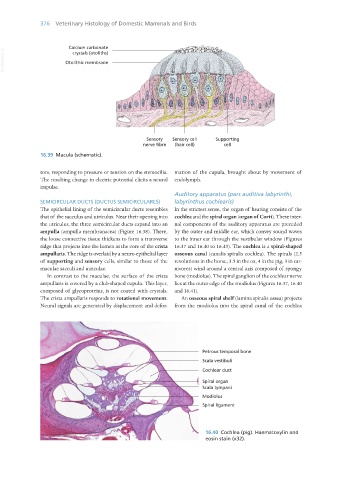
Auditory apparatus (pars auditiva labyrinthi,
SEMICIRCULAR DUCTS (DUCTUS SEMICIRCULARES) labyrinthus cochlearis)
The epithelial lining of the semicircular ducts resembles In the strictest sense, the organ of hearing consists of the
that of the sacculus and utriculus. Near their opening into cochlea and the spiral organ (organ of Corti). These inter-
the utriculus, the three semicircular ducts expand into an nal components of the auditory apparatus are preceded
ampulla (ampulla membranacea) (Figure 16.38). There, by the outer and middle ear, which convey sound waves
the loose connective tissue thickens to form a transverse to the inner ear through the vestibular window (Figures
ridge that projects into the lumen as the core of the crista 16.37 and 16.40 to 16.43). The cochlea is a spiral-shaped
ampullaris. The ridge is overlaid by a neuro-epithelial layer osseous canal (canalis spiralis cochlea). The spirals (2.5
of supporting and sensory cells, similar to those of the revolutions in the horse, 3.5 in the ox, 4 in the pig, 3 in car-
maculae sacculi and utriculae. nivores) wind around a central axis composed of spongy
In contrast to the maculae, the surface of the crista bone (modiolus). The spiral ganglion of the cochlear nerve
ampullaris is covered by a club-shaped cupula. This layer, lies at the outer edge of the modiolus (Figures 16.37, 16.40
composed of glycoproteins, is not coated with crystals. and 16.41).
The crista ampullaris responds to rotational movement. An osseous spiral shelf (lamina spiralis ossea) projects
Neural signals are generated by displacement and defor- from the modiolus into the spiral canal of the cochlea
16.40 Cochlea (pig). Haematoxylin and
eosin stain (x32).
Vet Histology.indb 376 16/07/2019 15:08