
Page 390 - Veterinary Histology of Domestic Mammals and Birds, 5th Edition
P. 390
372 Veterinary Histology of Domestic Mammals and Birds
VetBooks.ir
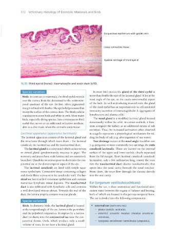
16.35 Third eyelid (horse). Haematoxylin and eosin stain (x70).
Species variation In most bird species the gland of the third eyelid is
Birds: In contrast to mammals, the third eyelid extends more than double the size of the lacrimal gland. It lies at the
over the cornea from the dorsonasal to the ventrotem- nasal angle of the eye, on the caudo-ventromedial aspect
poral quadrant of the eye. Its free, often pigmented of the bulb. As well as producing mucoid tears, the gland
margin is lined with feather-like epithelial processes that of the third eyelid has an important role in cell-mediated
sweep the surface of the cornea clean. The third eyelid is immunity (secretion of immunoglobulin A, aggregates of
translucent in some birds and white in owls. Most water lymphocytes and plasma cells).
birds, especially diving species, have a transparent third The nasal gland is a modified lacrimal gland located
eyelid that serves as an additional refractive medium, dorsonasally within the orbit. In certain seabirds, it func-
akin to a dive mask, when the animal is underwater. tions alongside the kidney as an additional means of salt
excretion. Thus, the increased lacrimation often observed
Lacrimal apparatus (apparatus lacrimalis) in seagulls represents a physiological mechanism for rid-
The lacrimal apparatus consists of the lacrimal gland and ding the body of salt (e.g. after ingestion of sea water).
the structures through which tears drain – the lacrimal Tear drainage occurs at the nasal angle via either one
canaliculi, the lacrimal sac and the nasolacrimal duct. (e.g. penguins) or more commonly two openings, the ostia
The lacrimal gland is a compound tubulo-acinar serous canaliculi lacrimalis. These are located on the internal
or mixed gland (predominantly mucous in pigs). The surface of the upper and lower eyelids, clearly separated
secretory end pieces have wide lumina and are extensively from the lid margin. Short lacrimal canaliculi (canaliculi
branched. Glandular secretions pass via ducts into the con- lacrimales), only a few millimetres long, convey the tears
junctival sac at the dorsotemporal aspect of the eye. into the nasolacrimal duct (ductus nasolacrimalis) that
The lacrimal canaliculi are lined with simple squa- opens into the nasal cavity beneath the nasal conchae.
mous epithelium. Connective tissue containing collagen From there, the tears flow through the choana directly
and elastic fibres is present in the canalicular wall. The lac- into the oral cavity.
rimal sac has stratified columnar epithelium and contains
numerous lymphatic tissue deposits. The nasolacrimal Ear (organum vestibulocochleare)
duct is also infiltrated with lymphatic cells and contains Within the ear, a close anatomical and functional asso-
a well-developed venous plexus. Towards the end of the ciation exists between the organs of balance and hearing,
duct, the lamina propria contains mucous glands. both of which are housed in the petrous temporal bone.
The ear is divided into the following components:
Species variation
Birds: In domestic birds, the lacrimal gland is located · external ear (auris externa):
at the temporal angle of the eye, between the periorbita − pinna (auricle, auricula),
and the palpebral conjunctiva. It empties by a narrow − external acoustic meatus (meatus acusticus
duct (or ducts) into the conjunctival sac near the con- externus),
junctival fornix. Owls, which produce only a small − tympanic membrane (membrana tympanica),
volume of tears, do not have a lacrimal gland.
Vet Histology.indb 372 16/07/2019 15:07