
Page 395 - Veterinary Histology of Domestic Mammals and Birds, 5th Edition
P. 395
Receptors and sense organs (organa sensuum) 377
(Figures 16.40 to 16.42), incompletely dividing its interior. thelium (usually simple squamous) and are filled with
VetBooks.ir Membranes extending from the osseous spiral lamina to perilymph.
the wall of the cochlea divide the spiral canal into three
The cochlear duct lies between the scala vestibuli
and scala tympani, accompanying these on their course
fluid-filled channels (Figures 16.40 and 16.41) termed the:
through the cochlea. It is connected proximally to the sac-
· scala vestibuli, cule by the ductus reunions. At the tip of the cochlea, the
· cochlear duct (ductus cochlearis) and cochlear duct ends in a blind sac. In contrast to the scala
· scala tympani. vestibuli and scala tympani, the cochlear duct contains
endolymph.
The scala vestibuli begins at the base of the stapes at the
vestibular window and extends through the spiral canal COCHLEAR DUCT (DUCTUS COCHLEARIS)
of the cochlea to its tip, where it is continuous with the The cochlear duct is a triangular spiral passage. At the apex
scala tympani at the helicotrema. The scala tympani passes of the triangle, the duct is connected to the free edge of
through the spiral canal to the cochlear (round) window the lamina spiralis ossea of the modiolus (Figures 16.40
at the base of the cochlea. Both scalae are lined with epi- and 16.41). The wall of the cochlear duct is divided into
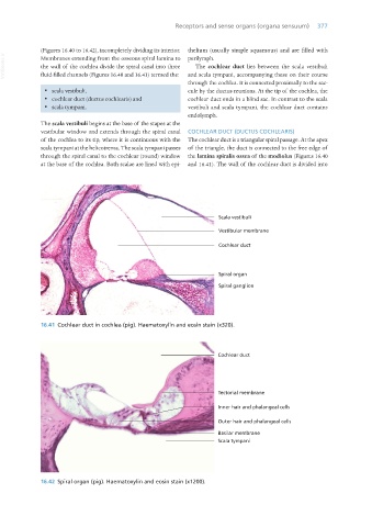
16.41 Cochlear duct in cochlea (pig). Haematoxylin and eosin stain (x320).
16.42 Spiral organ (pig). Haematoxylin and eosin stain (x1200).
Vet Histology.indb 377 16/07/2019 15:08