
Page 388 - Veterinary Histology of Domestic Mammals and Birds, 5th Edition
P. 388
370 Veterinary Histology of Domestic Mammals and Birds
ANTERIOR AND POSTERIOR CHAMBERS through the pupil into the anterior chamber (camera
VetBooks.ir nal chamber of the bulb bounded by the posterior surface anterior bulbi) from whence it passes, at the iridocorneal
The anterior chamber (camera anterior bulbi) is an inter-
angle, through the pectinate ligament into the spaces
of the cornea and the anterior surface of the iris. It com-
municates with the posterior chamber (camera posterior of Fontana of the cilioscleral sinus (Figure 16.14). From
bulbi) via the pupil (Figure 16.11 and 16.13). The walls of there it diffuses into the wide, usually bipartite scleral
the posterior chamber are formed by the posterior sur- venous sinus, which is located more superficially in
face of the iris, the ciliary body, the anterior surface of birds than in domestic mammals. In owls, the aqueous
the vitreous body and the lens. The anterior and posterior humour contains mucous substances secreted by the
chambers contain a clear watery fluid, aqueous humour, posterior corneal epithelium, making anterior chamber
containing electrolytes, glucose, amino acids and ascorbic paracentesis more difficult in these birds.
acid. The concentration of these substances in aqueous
humour differs from that in plasma. Accessory organs of the eye
Production of aqueous humour is a complex process in
which the blood–aqueous barrier (between the capillaries Eyelids (palpebrae)
and epithelium of the ciliary processes) plays an important The eyelids provide protection for the bulbs. In conjunc-
role. Aqueous humour is produced primarily by active tion with the precorneal tear film, they cleanse the anterior
+
transport of Na ions across the ciliary epithelium and by surface of the eye and prevent it from desiccating. The
osmotic transport of fluid into the posterior chamber. From palpebral reflex is an important component of this protec-
the posterior chamber, aqueous humour flows through the tive mechanism. In addition to an upper and lower eyelid
pupil into the anterior chamber. At the iridocorneal angle (palpebra superior and inferior), domestic mammals have
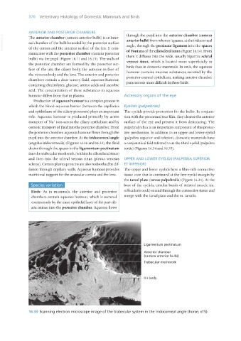
(angulus iridocornealis) (Figures 16.16 and 16.33), the fluid a conjunctival fold referred to as the third eyelid (palpebra
drains through the spaces in the ligamentum pectinatum tertia) (Figures 16.34 and 16.35).
into the trabecular meshwork (within the cilioscleral sinus)
and then into the scleral venous sinus (plexus venosus UPPER AND LOWER EYELIDS (PALPEBRA SUPERIOR
sclerae). Certain plasma proteins are also reabsorbed by dif- ET INFERIOR)
fusion through capillary walls. Aqueous humour provides The upper and lower eyelids have a fibre-rich connective
nutritional support for the avascular cornea and the lens. tissue core that is continued at the free eyelid margin by
the tarsal plate (tarsus palpebralis) (Figure 16.34). At the
Species variation base of the eyelids, circular bands of striated muscle (m.
Birds: As in mammals, the anterior and posterior orbicularis oculi) extend through the connective tissue and
chambers contain aqueous humour, which is secreted merge with the tarsal plate and the m. tarsalis.
continuously by the inner epithelial layer of the pars cili-
aris retinae into the posterior chamber. Aqueous flows
16.33 Scanning electron microscope image of the trabecular system in the iridocorneal angle (horse; x15).
Vet Histology.indb 370 16/07/2019 15:07