
Page 94 - Veterinary Histology of Domestic Mammals and Birds, 5th Edition
P. 94
76 Veterinary Histology of Domestic Mammals and Birds
VetBooks.ir
Microfibril
700 Å
2800 Å
Covalent cross-bonding and
quarter displacement of
collagen molecules within
collagen fibrils
2800 Å
15 Å Collagen molecule
– 120 AA 1052 AA Triple helix (2 a -peptide
1
chains and1 a -peptide
2
chain) of amino acids
of the pro-collagen
molecule with
terminal pro-peptides.
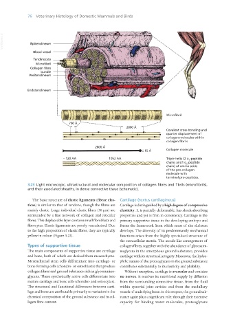
3.23 Light microscopic, ultrastructural and molecular composition of collagen fibres and fibrils (microfibrils),
and their associated sheaths, in dense connective tissue (schematic).
The basic structure of elastic ligaments (fibrae elas- Cartilage (textus cartilagineus)
ticae) is similar to that of tendons, though the fibres are Cartilage is distinguished by a high degree of compressive
mainly elastic. Large individual elastic fibres (30 μm) are elasticity. It is partially deformable, has shock-absorbing
surrounded by a fine network of collagen and reticular properties and yet is firm in consistency. Cartilage is the
fibres. This displaceable layer contains small fibroblasts and primary supportive tissue in the developing embryo and
fibrocytes. Elastic ligaments are poorly vascularised. Due forms the framework from which most of the skeleton
to the high proportion of elastic fibres, they are typically develops. The diversity of its predominantly mechanical
yellow in colour (Figure 3.22). functions arises from the highly specialised structure of
the extracellular matrix. The arcade-like arrangement of
Types of supportive tissue collagen fibres, together with the abundance of glycosami-
The main components of supportive tissue are cartilage noglycans in the amorphous ground substance, provides
and bone, both of which are derived from mesenchyme. cartilage with its structural integrity. Moreover, the hydro-
Mesenchymal stem cells differentiate into cartilage- or philic nature of the proteoglycans in the ground substance
bone-forming cells (chondro- or osteoblasts) that produce contributes substantially to its elasticity and pliability.
collagen fibres and ground substance rich in glycosamino- Without exception, cartilage is avascular and contains
glycans. These synthetically active cells differentiate into no nerves. It receives its nutritional supply by diffusion
mature cartilage and bone cells (chondro- and osteocytes). from the surrounding connective tissue, from the fluid
The structural and functional differences between carti- within synovial joint cavities and from the medullary
lage and bone are attributable primarily to variation in the vessels of underlying bone. In this respect, the ground sub-
chemical composition of the ground substance and in col- stance again plays a significant role; through their extensive
lagen fibre content. capacity for binding water molecules, proteoglycans
Vet Histology.indb 76 16/07/2019 14:56