
Page 89 - Veterinary Histology of Domestic Mammals and Birds, 5th Edition
P. 89
Connective and supportive tissues (textus connectivus) 71
Reticular connective tissue (textus connectivus Haemoreticular connective tissue
VetBooks.ir reticularis) (textus connectivus haemopoeticus)
Reticular connective tissue containing free blood cells,
Reticular connective tissue largely retains the character-
or their stem or progenitor cells, is referred to as haemo-
istics of undifferentiated mesenchyme. It is composed of
an open meshwork of reticular cells and delicate reticular reticular connective tissue (or haemopoietic tissue, e.g. in
fibres, as well as undifferentiated ground substance. bone marrow). Although it is a specialised tissue, it retains
Reticular cells usually have a large euchromatic nucleus its relationship with reticular tissue and may revert to this
that can increase in density based on the functional status tissue type or undergo conversion to adipose tissue (see
of the cell (nuclear pleomorphism). These cells synthesise Chapter 7, ‘Blood and haemopoiesis’).
reticular fibres and phagocytose dead cells and foreign par-
ticles. In addition, reticular cells are capable of recognising Adipose tissue (textus adiposus)
antigens on the cell surface and signalling this information Adipose tissue consists of a homogeneous population of
to immunocompetent cells. With its networks of closely fat cells (adipocytes) that develop from undifferentiated
interlinked reticular cells and fibres, reticular connective mesenchymal cells through the intracellular accumulation
tissue forms the structural framework of numerous organs of lipid droplets (Figures 3.16 to 3.19). Adipocytes occur
(e.g. lymphatic organs, liver, genital organs, subepithelial either individually or in groups (lobules), thus forming a
layers of the gastrointestinal tract). component of other tissues or organs. Adipose tissue has
an abundant blood supply.
Lymphoreticular connective tissue (textus The functions of adipose tissue are manifold. In the
connectivus lymphoreticularis) context of energy metabolism, it is capable of relatively
Lymphoreticular connective tissue (also referred to rapid storage and, as required, release of energy-rich
as lymphatic tissue) is reticular tissue in which the wide substrates. Preferential sites of fat storage include subcuta-
intercellular spaces have become populated with free neous tissue, the abdominal skin, the axilla and the inguinal
cells (macrophages, lymphocytes, plasma cells, mono- region.
cytes) (Figure 3.15). This tissue forms the stroma of Adipose tissue undergoes constant proliferation (triglyc-
lymphatic organs (lymph nodes, spleen, tonsils, thymus). eride synthesis, lipogenesis) and regression (triglyceride
Lymphoreticular tissue is a significant component of hydrolysis, lipolysis). Some of the fatty acids released
the adaptive and innate immune system (see Chapter 8, during lipolysis are re-esterified with glycerol 3 phosphate
‘Immune system and lymphatic organs’). (intracellular cycling).
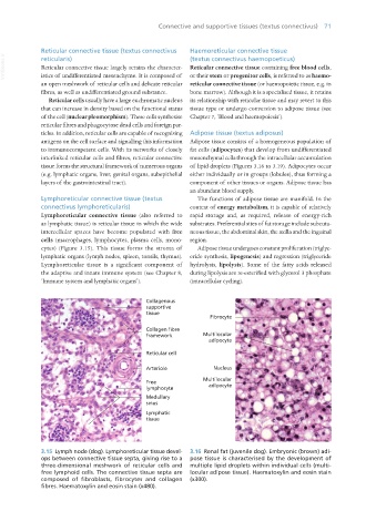
3.15 Lymph node (dog). Lymphoreticular tissue devel- 3.16 Renal fat (juvenile dog). Embryonic (brown) adi-
ops between connective tissue septa, giving rise to a pose tissue is characterised by the development of
three-dimensional meshwork of reticular cells and multiple lipid droplets within individual cells (multi-
free lymphoid cells. The connective tissue septa are locular adipose tissue). Haematoxylin and eosin stain
composed of fibroblasts, fibrocytes and collagen (x300).
fibres. Haematoxylin and eosin stain (x480).
Vet Histology.indb 71 16/07/2019 14:55