
Page 90 - Veterinary Histology of Domestic Mammals and Birds, 5th Edition
P. 90
72 Veterinary Histology of Domestic Mammals and Birds
Based on differences in structure and function, as well
VetBooks.ir as colour, location and vascularisation, adipose tissue
can be divided into:
· pluri- or multilocular adipose tissue (brown fat) and
· unilocular adipose tissue (white fat).
Pluri- or multilocular adipose tissue (textus
adiposus fuscus)
In pluri- or multilocular (brown) fat, the cytoplasm of
adipocytes contains numerous fat droplets of varying
size (Figures 3.16 and 3.18). This type of adipose tissue
develops from strands of cells containing large numbers of
mitochondria with abundant cytochrome (hence ‘brown’
fat). The individual adipocytes are smaller (15–25 μm) than
in white adipose tissue (see below), with a predominantly
centrally located nucleus and numerous glycogen and fat
vacuoles. Adrenergic nerve fibres extend to the cell surface,
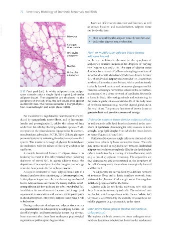
3.17 Foot pad (cat). In white adipose tissue, adipo-
cytes contain only a single lipid droplet (unilocular accompanied by a dense network of capillaries. Brown fat
adipose tissue). The organelles are displaced to the is found in birds, hibernating animals and rodents (e.g. in
periphery of the cell; thus, the cell boundaries appear the pectoral girdle). It also constitutes 5% of the body mass
as distinct lines. The nucleus occupies a marginal posi- of newborn mammals (e.g. near the thyroid gland and at
tion. Haematoxylin and eosin stain (x300).
the renal hilus). The primary functions of brown fat are to
generate heat and provide a source of energy.
Fat metabolism is regulated by neurotransmitters pro-
duced by sympathetic nerve fibres, and by hormones. Unilocular adipose tissue (textus adiposus albus)
Insulin and prostaglandin E inhibit the release of fatty In unilocular fat cells, lipid droplets deposited in the cyto-
1
acids from fat cells by blocking adenylate cyclase cAMP- plasm of lipoblasts (developing fat cells) coalesce to form
receptors on the plasmalemma (lipogenesis). In contrast, a single, large lipid droplet from which the tissue derives
noradrenaline, adrenaline, ACTH, TSH, GH and glucagon its name (Figures 3.17 and 3.19).
promote lipolysis by activating the adenylate cyclase cAMP Unilocular fat occurs as single cells or as clusters of cells
system. This results in cleavage of glycerol from triglycer- joined into lobules by loose connective tissue. The cells
ide molecules, with the release of free fatty acids into the may appear round or polyhedral (25–100 μm). Individual
capillaries. adipocytes are almost completely filled by the lipid droplet
A notable functional feature of adipose tissue is its (which is stabilised by a coating of microfilaments), with
tendency to revert to less differentiated tissue (following only a rim of cytoplasm remaining. The organelles are
depletion of stored fat). In ageing adipose tissue, the thus displaced to, and concentrated at, the periphery of
deposition of mucopolysaccharides can give rise to large the cell. Consequently, the nucleus is marginally located
vesicular, honeycomb-like fat cells (serous fat). and flattened.
As a poor conductor of heat, adipose tissue acts as a The adipocytes are surrounded by a delicate network
thermal insulator thus contributing to thermoregulation. of reticular fibres and a dense capillary network. Fine
It also plays an important role in withstanding mechanical periarteriolar plexuses of adrenergic nerve fibres regulate
forces. Adipose tissue surrounds the kidney and has a cush- metabolic processes within the tissue.
ioning effect in the foot pads and the orbit (retrobulbar fat). Adipose cells do not divide. However, new cells can
In addition, fat contributes to the structural integrity of form from other mesenchymal cells. The colour of uni-
organs and, in association with various joints, participates locular fat, which ranges from white (hence ‘white fat’)
in shock absorption. Moreover, adipose tissue plays a role to yellow, is determined by the quantity of exogenous fat-
in hydration. soluble pigments (e.g. carotenoids) in the tissue.
During embryonic development, adipose tissue serves
as a placeholder for subsequently developing tissues. Fat Connective tissue proper (textus connectivus
also fills lympho- and haemoreticular tissues (e.g. thymus, collagenosus)
bone marrow) after these have undergone physiological Throughout the body, connective tissue undergoes struc-
regression or pathological degeneration. tural and functional adaptations, based on the mechanical
Vet Histology.indb 72 16/07/2019 14:55