
Page 236 - Zoo Animal Learning and Training
P. 236
220 Tasks for the Veterinary Assistant
money, and may endanger an animal’s life. Make it your
mission to ensure all test results are written down!
Reflection
What technique or key thought will you use to
remember to write in the laboratory log book?
Learning Exercise
Develop a “normals” reference chart, keep it
pocket size so you have it with you at all times.
Maintenance of the
Common Laboratory
Equipment in the
Veterinary Lab
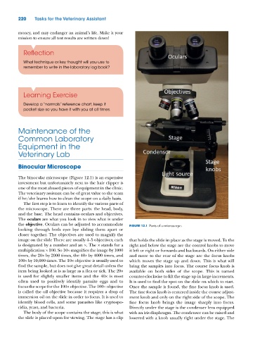
Binocular Microscope
The binocular microscope (Figure 12.1) is an expensive
investment but unfortunately next to the hair clipper is
one of the most abused pieces of equipment in the clinic.
The veterinary assistant can be of great value to the team
if he/she learns how to clean the scope on a daily basis.
The first step is to learn to identify the various parts of
the microscope. There are three parts: the head, body,
and the base. The head contains oculars and objectives.
The oculars are what you look in to view what is under
the objective. Oculars can be adjusted to accommodate FIGURE 12.1 Parts of a microscope.
looking through both eyes bye sliding them apart or
closer together. The objectives are used to magnify the
image on the slide There are usually 4–5 objectives; each that holds the slide in place as the stage is moved. To the
is designated by a number and an ×. The × stands for a right and below the stage are the control knobs to move
multiplication × 100. So 10× magnifies the image by 1000 it left or right or forwards and backwards. On either side
times, the 20× by 2000 times, the 40× by 4000 times, and and more to the rear of the stage are the focus knobs
100× by 10,000 times. The 10× objective is usually used to which moves the stage up and down. This is what will
find the sample, but does not give great detail unless the bring the samples into focus. The course focus knob is
item being looked at is as large as a flea or tick. The 20× available on both sides of the scope. This is turned
is used for slightly smaller items and the 40× is most counter‐clockwise to lift the stage up in large increments.
often used to positively identify parasite eggs and to It is used to find the spot on the slide on which to start.
focus the scope for the 100× objective. The 100× objective Once the sample is found, the fine focus knob is used.
is called the oil objective because it requires a drop of The fine focus knob is centered inside the course adjust-
immersion oil on the slide in order to focus. It is used to ment knob and only on the right side of the scope. The
identify blood cells, and some parasites like cryptospo- fine focus knob brings the image sharply into focus.
ridia, yeast, and bacteria. Directly under the stage is the condenser lens equipped
The body of the scope contains the stage; this is what with an iris diaphragm. The condenser can be raised and
the slide is placed upon for viewing. The stage has a clip lowered with a knob usually right under the stage. The