
Page 241 - Zoo Animal Learning and Training
P. 241
Chapter 12 Laboratory Skills 225
(a) (b)
FIGURE 12.6 (a) Demodex and (b) burrowing mite species. Source: (a) Wikimedia Commons. Used under CC BY‐SA 3.0, https://
commons.m.wikimedia.org/wiki/File:Haarbalgmilbe.jpg (b) Source: Wikimedia Commons. Used under CC BY-SA 2.5, https://
commons.m.wikimedia.org/wiki/File:Canine_scabies_mite.JPG.
Mosquitoes
In addition to being irritants and voracious bloodsuckers,
mosquitoes are most noted for their transmission of dis-
eases such as Dirofilaria immitus (heartworms) and West
Nile disease, which is zoonotic. Mosquitoes require water
to lay their eggs and different species use different pools of
water. They are more active in the evening and early morn-
ing hours. Topical and oral medications and vaccines are
used as preventatives for the diseases they transmit.
Mites
Mites feed on tissue fluids and skin cells. They can cause
severe dermatitis accompanied by pruritus and alopecia.
Burrowing mites such as Demodex, Notoedres, and Sarcoptes
require a skin scraping to find (Figure 12.6). The
supplies needed for this test are a scalpel blade, a micro-
scope slide with a drop of mineral oil upon it, and a
microscope. The veterinarian or technician will scrape
the area in question with the scalpel blade and deposit
the material onto the slide. The Demodex are cigar‐shaped
and the others have eight short legs on rounded bodies.
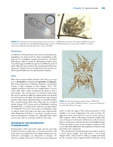
The non‐burrowing mites have long legs on rounded FIGURE 12.7 Non‐burrowing mite species. Source: Wikimedia
bodies (Figure 12.7). Some, such as Cheyletiella (“walking Commons. Used under CC BY‐SA 3.0, https://commons.m.wikimedia.
org/wiki/File:Otodectes‐mite.jpg.
dandruff mite”) is visible to the naked eye as white flakes
of “dandruff” that move! Use a piece of cellophane tape
to capture them, then place the tape onto a microscope order to find the eggs of the internal parasites. If left
slide. Diagnosis of Otodectes (“ear mites”) requires an ear outside or at room temperature for even an hour the
swab as discussed in Chapter 10, although these mites eggs may hatch and therefore not be found giving a
can be seen with an otoscope inside the ear canal. false negative. When collecting a voluntary sample take
note of shape; is it formed, semi‐formed, soft or watery?
Sampling for and Identification Is there any mucus or blood, are there any visible worms?
of Endoparasites These are things to note on the sample container as the
act of collection will distort these physical attributes that
Endoparasites infect internal organ systems and may may help with a diagnosis.
result in diarrhea, weight loss, or anemia; therefore, the On occasion the veterinarian may not want to wait for
fecal exam is an important and common laboratory test. a freely given sample and will utilize a fecal loop
Collection techniques are discussed in Chapter 10. (Figure 12.8). This is used to collect a sample internally
However, samples need to be fresh out of the patient in via the rectum. Items required will be lubricant, fecal