Page 242 - Zoo Animal Learning and Training
P. 242
226    Tasks for the Veterinary Assistant


                                                             to the top of a test tube which is captured on a coverslip
                                                             and then transferred to a microscope slide. There are two
                                                             standard techniques: a float or a centrifuge preparation.

                                                             Fecal Float Technique

                                                             Equipment required: 10–12 mL test tube, test tube rack,
                                                             microscope slide, coverslip, two paper cups, strainer, two
                                                             applicator sticks, and fecal solution.
                                                               Preparation:
                                                             1.  Using the applicator sticks, place a “quarter” sized
                                                                piece of feces into one of the paper cups and add
                                                                approximately 20–30 mL of fecal solution. You can use
                                                                the test tube to measure out the fecal solution. Place
                                                                the test tube into the test tube rack for future use.
                                                             2.  Mix the feces and fecal solution thoroughly with the
                                                                sticks. Place the strainer over the second cup and
                                                                pour the fecal mixture through the strainer. Set the
                                                                strainer and first cup into the sink.
                                                             3.  Pour the strained fecal mixture into the test tube
                                                                until a meniscus forms. Don’t overfill as this may
                                                                wash the parasite eggs out of the sample.
                                                             4.  Carefully place a coverslip on top of the test tube and
                                                                set a timer for 10–15 minutes. Alert the technician
                                                                that the time is set.


                                                             Centrifuge Technique
                                                             The same equipment is required as for the flotation
                                                             method.
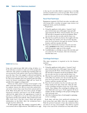
          FIGURE 12.8  Fecal loop.
                                                               Procedure:
                                                             1.  Using the applicator sticks, place a “quarter” sized
          loop, and a microscope slide with a drop of saline or a   piece of feces into one of the paper cups and add
          fecal collection container to take care of the sample after   approximately 20–30 mL of fecal solution. You can use
          collection. The patient is usually kept standing with one   the test tube to measure out the fecal solution. Place
          arm around the neck and the other hand to hold the tail   the test tube into the test tube rack for future use.
          up at the base. Once the sample is collected and labeled   2.  Mix the feces and fecal solution thoroughly with the
          properly, alert the technician that a sample is waiting. If   sticks. Place the strainer over the second cup and
          he/she is not available, place the sample in the lab refrig-  pour the fecal mixture through the strainer. Set the
          erator for future examination. Eggs will not hatch as   strainer and first cup into the sink.
          quickly if the sample is refrigerated.             3.  Pour the fecal solution into the test tube up to the
            Clients are often asked to bring a fecal sample to the
          clinic at the time of their appointment or to drop one off   10 mL mark on the side of the tube. Take the tube
                                                                to the centrifuge and place it into one of the tubes
          for analysis. Instruct the client to watch the animal defe-  inside. Then balance the centrifuge by adding a test
          cate to be certain it has come from their animal and it is   tube filled with water up to 10 mL opposite the fecal
          fresh. Have them place the feces into a clean, air‐tight,   sample. Set the centrifuge at the proper settings and
          leak‐proof container and keep it cool. If the sample is   time and push start.
          collected the night before the appointment it must be   4.  Alert the technician that a fecal sample is spinning
          placed in a refrigerator or in an ice chest otherwise the   in the centrifuge.
          eggs hatch and the larva will die. When delivered to the
          clinic make sure it has the correct client and patient   Each clinic will have their preferred technique for
          information on the label. Alert the technician that a   fecal set‐up that may differ from the examples given.
          sample has been delivered.                         Find the protocol manual or have the technician show
            To find parasite eggs, the sample is prepared with a   you how to set up a fecal sample. This will be of great
          concentrated salt or sugar solution. This will float the eggs   help to the team if the technicians are busy with clients.

