
Page 120 - parasitology for medical and clinical laboratoryprofessionals
P. 120
100 CHAPTER 4
Wuchereria bancrofti, Brugia malayi, and B. timori, which
are transmitted by mosquitoes. The discovery of the life
cycle by a Scotsman, Patrick Manson, in 1877 is regarded
as one of the most significant discoveries in tropical medi-
cine, but in the context of the history of parasitology it is
better perceived as a logical extension of much that had
gone before. Like Dracunculus, the adult filarial worms Source: Centers for Disease Control and Prevention (CDC)
live in subcutaneous tissues, but unlike Dracunculus, the
larvae, called microfilariae, produced by the female worm
pass into the blood and are taken up by a bloodsucking
mosquito when it feeds. After development in the mos-
quito, the microfilariae are injected into a new host upon
which the mosquito feeds again. The microfilaria, when
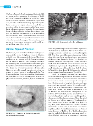
present in the circulatory system, are readily visible in a FIGURE 4-18 Elephantiasis of leg due to filariasis
stained blood smear (Figure 4-17).
Clinical Signs of Filariasis limbs and genitalia may have been the artists’ expressions
of creativity in at least some of the ancient artistic ren-
Elephantiasis, in which the lower limbs are misshapen and derings. Lymphatic filariasis has been found along the
appear similar to that of elephants with little definition Nile River from past evidence as well as current victims
and with loose skin, was a particular form of the disease of the infection. Some prominent figures from ancient
that has been met with a great deal of attention through- civilizations show the swollen limbs of a victim of micro-
out the history of mankind. This grotesque swelling of filariasis. The statue of the Egyptian Pharaoh Mentuho-
the limbs may also include that of breasts and the geni- tep II from about 2000 BC shows evidence of possible
tals, particularly for the scrotum of men (Figure 4-18). elephantiasis in grotesquely enlarged limbs. In addition,
The disease was obviously present since before recorded small statuettes and gold weights from the Nok culture
history, as artwork from early man shows drawings and in West Africa from about AD 500 depict the enlarged
statues of persons who may have been suffering from scrota characteristic of elephantiasis (Cox, 2002).
lymphatic filariasis. However, some of the drawings were Greek and Roman writers as well as Arabic physi-
highly stylistic and included exaggerations of certain cians were careful to point out the differences between
physical characteristics, therefore, depictions of swollen the physical effects of leprosy and those of elephantiasis
from infection with the microfilaria (small worms). The
first definitive reports of lymphatic filariasis only began
to appear in the sixteenth century. The condition of fi-
lariasis was so well known that the common name “the
Source: Centers for Disease Control and Prevention (CDC) between 1588 and 1592, and the incident was docu-
curse of St. Thomas,” was named after those who killed
St. Thomas. His death occurred during a visit to Goa
mented when a Dutch explorer named Jan Huygen Lins-
choten recorded that the descendants of those that killed
St. Thomas were “all born with one of their legs and one
foot from the knee downwards as thick as an elephant’s
leg” (Cox, 2002). References to the disease of filariasis
ing China, where Patrick Manson’s studies in 1877 led
to the discovery of the life cycle of the filarial parasites.
Another pathological condition associated with lym-
FIGURE 4-17 Posterior of Wuchereria bancrofti are available in other areas of Africa and Asia, includ-
microfilaria in blood smear phatic filariasis is chyluria, in which the urine appears