
Page 124 - parasitology for medical and clinical laboratoryprofessionals
P. 124
104 CHAPTER 4
of the mosquito the larvae grow and develop into an in-
fective stage over a period of perhaps 10 days. The infec- MICROSCOPIC DIAGNOSTIC
tive larvae, which range from 1 to 2 mm long, then move FEATURE
to the proboscis of the mosquito and during the next
blood meal the insect infects the next host the mosquito
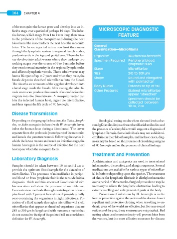
General
bites. The larvae injected into a new host then move
Classification—Microfilaria
through the lymphatic system to regional lymph nodes,
predominantly in the legs and genital area. There the lar- Organism Wuchereria bancrofti
vae develop into adult worms where they undergo two Specimen Required Peripheral blood,
molting stages over the course of 6 to 9 months before lymphatic fluid
they reach sexual maturity in the regional lymph nodes Stage Microfilariae
and afferent lymphatic vessels. These adult worms may
Size 245 to 300 μm
have a life span of up to 7 years and when they mate, the
Shape Round and elongated
female deposits sheathed microfilariae into the blood.
with pointed tail
The sheaths are remnants of the egg that developed into
Body Nuclei Extends to tip of tail
a larval stage inside the female. After mating, the adult fe-
Other Features Stained microfilariae
male worm can produce thousands of microfilariae that
appear “sheathed”
migrate into the bloodstream. A mosquito vector can
Specimen should be
bite the infected human host, ingest the microfilariae,
collected between
and thus repeat the life cycle of W. bancrofti.
10 PM, 2 PM
Disease Transmission
Depending on the geographic location, the Culex, Anoph- Serological testing results where elevated levels of se-
eles, or Aedes mosquito infected with W. bancrofti larvae rum IgE (antibodies) as elevated antifilarial antibodies and
infect the human host during a blood meal. The larvae the presence of eosinophilia would support a diagnosis of
separate from the proboscis (mouthpart) of the mosquito lymphatic filariasis. Some individuals may not exhibit mi-
and invade the puncture wound. Following the cycles in crofilariae in their blood samples, and in these cases, diag-
which the larvae mature and reach an infective stage, the nosis may be based on the presence of circulating antigens
human host again is the source of infection for the next of W. bancrofti and on the presence of clinical findings.
host upon which the mosquito feeds.
Treatment and Prevention
Laboratory Diagnosis
Antihistamines and analgesics are used to treat related
Samples should be taken between 10 pm and 2 am to inflammation, discomfort, and allergic responses. Several
provide the optimum blood sample for the detection of medications are available for various types of microfilar-
microfilariae. The presence of microfilariae in periph- ial infections depending upon the species. The treatment
eral blood or from lymphatic fluid is the most definitive of choice for lymphatic filariasis is diethylcarbamazine
diagnosis. Thick and thin smears of blood stained with over a period of three weeks. Surgical procedures may be
Giemsa stain will show the presence of microfilariae. necessary to relieve the lymphatic obstruction leading to
Concentration methods through centrifugation of sam- extreme swelling and enlargement of parts of the body.
ples fixed with 2 percent formalin will provide a buffy Prevention of infections by W. bancrofti is in the
coat containing the organisms in light infections. Fil- form of protection against the vectors of the disease. Insect
tration of a fluid sample through a microfilter will yield repellent and protective clothing when travelling to en-
microfilariae that appear as sheathed organisms that are demic areas of the world are effective, but for year-round
245 to 300 μm in length and with numerous nuclei that residents of the area, these measures are not practical. Bed
do not extend to the tip of the pointed tail are considered netting when used conscientiously will prevent bites from
definitive for W. bancrofti. the vectors, but the most effective measures for disease