
Page 72 - Tobillo y Pie 9.1
P. 72
Managing the adult flexible flatfoot deformity. The past, present and the future. An evolution of thinking
A B
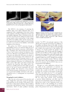
Figure 2. The pre and postoperative radiographs of an obese
female patient with a rupture of the PTT and a rupture of the
spring ligament. This was treated with a subtalar arthrodesis,
transfer of the FDL, and a spring ligament reconstruction
The MDCO is the mainstay of correcting the
hindfoot valgus, and is either performed alone, or in C
conjunction with a lengthening of the lateral column Figure 3. Calcaneal osteotomy. The skin is retracted (A), the
of the calcaneus, and aims to change the calcaneal axis osteotomy performed and opened with a laminar spreader
and hindfoot alignment. This in turn helps protect the (B) and a guide pin inserted for a cannulated screw or a
soft tissue reconstruction by taking the tension off the specific calcaneus medial displacement plate following
tendon transfer or the reconstruction. It also realigns 10mm of medial displacement (C)
the pull of the Achilles tendon and the moment arm
of the gastrocnemius soleus complex is converted from
an everter to an inverter of the hindfoot with medial eversion. The PTT is the principle supinator of the
translation of the calcaneus. subtalar joint along with functioning as an adductor of
the midfoot and plantar flexor of the ankle. So if this
We perform the MDCO osteotomy through a tendon is ruptured, we have to replace it with something
lateral oblique incision, one centimeter below the tip of else to balance the muscle forces of the hindfoot, but
the fibula in line with the osteotomy. A full thickness in doing so we should consider the strength of the
flap is developed with subperiosteal dissection down transferred muscle. The FDL has only 28% strength of
to bone. Care is taken to protect the branches of the the PTT and the FHL has 50% strength of the PTT.
sural nerve, although we warn patients that numbness Although some surgeons have advocated transfer of the
is frequent post operatively. Retractors are placed on FHL instead of the FDL to replace the torn PTT, 100%
the plantar and dorsal aspects of the calcaneus for soft of patients report loss of FHL strength and we believe
tissue protection and a self-retaining retractor is also this is not an acceptable outcome. However, transfer
used to stretch the margins of the incision and maintain of the FDL alone is also not adequate and the medial
exposure during the osteotomy. An oscillating fan saw shift of the calcaneus with the MDCO does not ever
blade is used at right angle to the lateral calcaneal wall compensate for this imbalance.
to perform the osteotomy. The medial wall of the
calcaneus is carefully perforated with a slight punching Although our earlier publications indicated that
action of the saw to prevent inadvertent soft tissue the results of this combination of treatment i.e.
damage medially. After distraction of the osteotomy FDL transfer with MDCO was satisfactory, we now
with a laminar spreader, a displacement of 10 to 12mm recognize that a transfer of the FDL is not the ideal
can be performed medially and fixation with either a procedure for correction of the balance of deformity.
cannulated screw or a locking plate can be performed The FDL is far weaker than the PTT, and regardless
(Figure 3). of the additional procedures performed to improve the
structure of the foot, the muscle imbalance remains.
Managing the muscle imbalance We also noted many patients slowly developing
This is the key to the success of the procedure and a recurrent flatfoot deformity, and while many of
many changes have taken place in my own approach these were not symptomatic, given the deformity, we
to the problem over the decades. With rupture of the believed that sooner or later symptoms would recur.
PTT, there is always muscle imbalance due to weakness Therefore we have to do something else to increase
of inversion, and unbalanced activity of the peroneal inversion power, or to consider weakening the eversion
musculature, which of course produces increasing power. The peroneus brevis acts as a deforming force
62 Tobillo y Pie 2017;9(1):58-68