
Page 77 - Tobillo y Pie 9.1
P. 77
Myerson M, Li SY
of the posterior facet. One must always therefore open
the capsule over the subtalar joint and ensure that this
impingement is not present. If the impingement is
noted, which in our experience is very common, then
a saw must be used to shave down the bone next to
A B the posterior facet until the range of motion no longer
causes impingement.
The procedure is performed by retracting the peroneal
tendons and marking the osteotomy with a k-wire 1cm
proximal to the joint (Figure 10). The osteotomy is
made with a saw, cutting the width of the calcaneus from
C D lateral to medial, it is then distracted, and the size of the
graft determined under fluoroscopy to ensure that good
coverage of the talonavicular joint has been obtained.
This is attained using a pin distractor specifically designed
for this procedure. In selected cases the peroneus brevis
tendon is then transferred to the peroneus longus tendon
E F to decrease the abduction and eversion force on the
hindfoot.
We believe that function of the foot will improve if
the shape of the foot is completely corrected, i.e “function
follows form” and for this reason are now routinely
performing an opening wedge osteotomy of the medial
G H cuneiform (the Cotton procedure) or an arthrodesis
st
of the 1 TMT joint, even for cases where the forefoot
supination is minimal. Although this is not done strictly
according to our classification above, the addition of
this osteotomy seems to improve the alignment of all
the feet irrespective of the extent of forefoot supination.
In addition, we have noted that by using the cuneiform
I osteotomy, there is far less need for an arthrodesis of either
st
the 1 tarsometatarsal joint or the naviculocuneiform
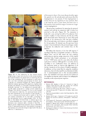
Figure 10. A) The osteotomy for the lateral column joint. The cuneiform osteotomy increases the tension on
lengthening is made 1cm proximal to the calcaneocuboid the windlass mechanism, and in doing so the radiographic
joint and the location is marked with a k-wire to guide the instability at the adjacent joints improves.
axis of the osteotomy; B) The osteotomy is made with a
saw, and then distracted approximately 8-10mm using
a specific pin distractor (Paragon 28, Denver Colorado). REFERENCES
Note the biplanar opening of the osteotomy, slightly wider 1. Myerson MS, Badekas A, Schon LC. Treatment of Stage II
dorsally and laterally; C) The graft is inserted and the pin posterior tibial deficiency with FDL transfer and calcaneal
distractor removed. D) The osteotomy is secured with a osteotomy. Foot Ankle Int. 2004; 25(7):445-50.
specific plate designed for a lateral column lengthening 2. Johnson K.A. PTT rupture. Clin Orthop. 1983;(177):140-7.
(Paragon 28, Denver Colorado); E) Due to the severity 3. Johnson KA, Strom DE. PTT dysfunction. Clin Orthop. 1989; (239):
of the abduction deformity, the peroneus brevis tendon 196-206.
was then transferred to the peroneus longus tendon; F) 4. Mann RA. Acquired flatfoot in adults. Clin Orthop Relat Res. 1983;
This is a 54-year-old female with a rupture of the PTT, (181):46-51.
and marked abduction of the foot with uncovering of 5. Myerson MS,Corrigan J. Treatment of PTT dysfunction with FDL
the talonavicular joint of approximately 50%; G) Note transfer and calcaneus osteotomy. Orthopaedics. 1993;19(5):
383-8.
also on the lateral XR that there is marked sag at the 6. Koutsougiannis EJ. Treatment of mobile flatfoot by osteotomy of
1 tarsometatarsal joint, which requires correction; The the calcaneus. J Bone Joint Surg Br. 1971;53(1):96-100.
st
deformity was corrected with a transfer of the FDL, a 7. Haddad SL, Myerson MS, Younger A, Anderson RB, Davis WH,
lateral column lengthening osteotomy and an arthrodesis Manoli A 2nd. Symposium: Adult acquired flatfoot deformity.
of the 1 tarsometatarsal joint. Note excellent coverage of Foot Ankle Int. 2011;32(1):95-111.
st
the talonavicular joint (H) and good alignment of the talus 8. Monteagudo M, Maceira E. Posterior tibial tendoscopy. Foot Ankle
st
with the 1 metatarsal (I). Clin. 2015;20(1):1-13.
Tobillo y Pie 2017;9(1):58-68 67