
Page 74 - Tobillo y Pie 9.1
P. 74
Managing the adult flexible flatfoot deformity. The past, present and the future. An evolution of thinking
sustentaculum. A lateral radiograph must be obtained
to verify that it is not too close to the subtalar joint or
the edge of the sustentaculum which can cause fracture.
The 4.5mm drill hole is then made and the allograft
tendon is inserted into the hole with an interference
screw or suture anchor holding the position securely. The
second hole is made from the plantar medial inferior
pole of the navicular aiming dorsally out the center of
the navicular. The tendon is then pulled through and
an interference screw is inserted under the navicular to
maintain maximum tension (Figure 5). The tension is
Figure 6. Radiographic appearance before and after
reconstruction with a spring ligament repair. In this
case, there was a rupture of the spring ligament with
an associated flatfoot but the PTT was quite normal
Figure 5. The FDL is visualized passing from inferior with no tenosynovitis nor rupture
to superiorly through the bony tunnel created in the
navicular. It is sutured to the stump of the PTT and then
dorsally to the periosteum over the navicular
set with the foot in slight varus at the talonavicular joint.
If this procedure is performed in conjunction with an
FDL transfer then one has to be careful with the drill
tunnels in the navicular to prevent fracture. There are
times when the spring ligament is stretched out, but
the pathology of the capsuloligamentous pathology
extends to the deltoid ligament as well. In these cases,
the reconstruction is performed using a graft extending
from the medial malleolus to the navicular as above.
Both of these graft procedures can be reinforced with
heavy braided sutures which are available commercially
(the suture brace, Arthrex, Naples Florida). However
the suture brace is extremely rigid and one has to be
careful with the tension that is set on the medial ankle
to prevent overcorrection. Furthermore, the suture
brace cannot substitute for capsuloligamentous tissue,
and must be applied on the top of this tissue and never
inserted as an intra-articular suture.
As mentioned previously the FDL is a weaker
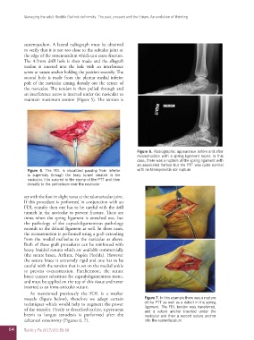
muscle (figure below), therefore we adopt certain Figure 7. In this example there was a rupture
techniques which would help to augment the power of the PTT as well as a defect in the spring
ligament. The FDL tendon was transferred,
of the transfer. Firstly as described earlier, a peroneus and a suture anchor inserted under the
brevis to longus tenodesis is performed after the navicular and then a second suture anchor
calcaneal osteotomy (Figures 6, 7). into the sustentaculum
64 Tobillo y Pie 2017;9(1):58-68