
Page 75 - Tobillo y Pie 9.1
P. 75
Myerson M, Li SY
The future of management of the rupture resulting in fatty infiltration of the muscle and
adult acquired flatfoot deformity permanent loss of function (Figure 8). Secondly, even
It is always difficult to predict where we are going
with management of the adult acquired flatfoot
deformity. Certainly, as indicated above, muscles’
balancing is critical, and we have approached this with a
release of the deforming peroneus brevis and transfer to
the peroneus longus in order to improve the balance
of forces of the hindfoot. This is the one additional
procedure that we would recommend routinely. The
next generation of management of PTT reconstruction
will involve a better understanding of the use of the
PT muscle without sacrificing it’s power. There are two
ways in which this can be accomplished. The first is
to preserve the tendon, despite the rupture, perform a
repair of the tendon and then perform the FDL transfer.
As we noted in the introduction, living collagen cells
remain present in the ruptured tendon indicating the Figure 8. Note the fatty atrophy of the PT
potential for healing. Undoubtedly the reason that this muscle in this leg. This is a contraindication to
procedure failed in the early 1980’s was that the rest of performing an allograft tendon reconstruction
the foot was ignored and the forces of the torn PTT
were not treated. While this is not a procedure that we
routinely perform, where do you begin with excision
of the torn PTT, and where do you perform a repair?
Assuming that the pain from the torn tendon is the
result of abnormal forces on the tendon, then various
osteotomies will realign the foot, and an endoscopy
of the PTT may be quite sufficient to evaluate the
PTT, perform a limited debridement or guide one to
opening the tendon. Endoscopy of the tendon has a
(8)
role, but cannot be used unless additional procedures A
are performed to ensure a plantigrade foot.
We have to recognize that if the PTT is ruptured,
the PT muscle may still however be functioning. To
avoid the problems that I note above with the muscle
imbalance, surely it would be preferable to use a tendon
graft to replace the torn PTT instead of the FDL
transfer? In this way, you are able to preserve the power
of the posterior tibial muscle. It should be understood
however that if the PTT graft procedure is used, then
it should not be necessary to add a peroneus brevis
to longus tenodesis. This procedure is discussed in
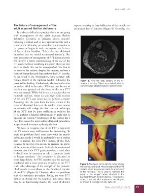
more detail below. An FDL transfer into the navicular B
combined with a more proximal tenodesis to the PTT Figure 9. The figure on the left (A) demonstrates
could take advantage of the strength of the posterior the two incisions used. Note the stump of the PTT
tibial muscle to contribute to the functional transfer distally, which is preserved with its attachment
of the FDL (Figure 9). However, there are problems to the navicular. This is used to reinforce the
with this tenodesis procedure. Firstly, the torn PTT repair of the allograft when attached to the
navicular through the bony tunnel. The proximal
cannot or should not be routinely used since there stump is sutured with a weave to the allograft
may be no functioning muscle, the result of chronic as shown in the figure on the right (B)
Tobillo y Pie 2017;9(1):58-68 65