
Page 73 - Tobillo y Pie 9.1
P. 73
Myerson M, Li SY
in the flatfoot deformity and the unopposed pull of We transfer the FDL to the navicular by drilling
the peroneus brevis causes eventual elongation of the through the bone with a 4.5mm drill and passing the
medial supporting structures, eventually leading to an tendon through the intraosseous tunnel from plantar to
abduction deformity. In the absence of a functioning dorsal. The tendon is then tensioned and sutured onto
PTT, the peroneus brevis therefore will always contribute the periosteum both on the superior and undersurface
to the worsening of the flatfoot deformity. Therefore of the navicular. It is not necessary to use an interference
we now recommend a transfer of the peroneus brevis screw, and this suture repair allows immediate weight
tendon to the peroneus longus in conjunction with the bearing without concern for stretching. The tension
FDL transfer, and the calcaneus osteotomy. By virtue that is set on the tendon during suture is important. The
of the insertion of the peroneus longus on the base of tendon must be tight, and although there are different
the 1 metatarsocuneiform joint the tendon transfer opinions as to just how tight this transfer should
st
helps to strengthen the first metatarsal plantarflexion be, my preference is to tighten the tendon slightly to
and reduces slightly the eversion of the hindfoot produce very slight inversion of the foot. This should
thereby improving the deformity correction. This can be half way between maximum tension and maximum
be performed through the same incision used for the relaxation. Some surgeons place the tendon tension
calcaneal osteotomy by extending it slightly proximally at maximum but this cannot possibly be the correct
and distally. tension to apply to any tendon transfer.
Managing the spring ligament tear
Flexor digitorum longus (FDL) transfer
surgical technique The function of the spring ligament is to maintain
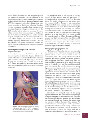
A medial incision over the PTT is made and the the position of the talar head, forming the medial
plantar sector of the articular cavity known as
tendon inspected to determine if just a debridement “acetabulum pedis”. The spring (calcaneo navicular)
will be sufficient or whether an FDL transfer or tendon ligament, the deltoid ligament, the plantar ligaments
graft procedure is indicated, depending on the disease and the plantar fascia in a passive way with the
staging. It is not always easy to see the rupture of the posterior tibial tendon in an active way, function to
PTT tendon, but by rotating the tendon the tear is seen stabilize the subtalar joint and the medial longitudinal
since it is usually on the posterior surface of the tendon arch. It is essential that one always inspect the spring
(Figure 4). ligament complex during repair and reconstruction of
the ruptured PTT. Furthermore, injury of the spring
ligament can occur in isolation not associated with a
tear of the PTT. While this isolated injury of the spring
ligament is not common it does occur, and we have
frequently made the error of assuming that a rupture of
the PTT is present when it is the torn spring ligament
that produces the exact same deformity of the hindfoot.
The clinical diagnosis of this isolated tear is not easy,
because the patient will have pain at the insertion of
A the PTT, but normal power and strength of the PTT
is present. Eventually, rupture of the spring ligament
will lead to a more vertically oriented talus.
Historically, we attempted repair of the spring
ligament with sutures, but these are rarely strong enough
to support the repair. An alternative treatment is to place
one suture anchor into the navicular and another into
the sustentaculum tali and then use the sutures from
the anchors to reinforce the repair of the ligament. As
B an alternative, we occasionally use a tendon graft which
passes from the sustentaculum tali through a tunnel under
Figure 4. Note the tenosynovitis and the tearing
and narrowing of the PTT (A). The rupture of the plantar medial head of the navicular to support the
the tendon is frequently visible on the posterior head of the talus. A 4.5mm drill hole is made over a
surface when rotating the tendon (B) cannulated guide pin which is inserted 1cm under the
Tobillo y Pie 2017;9(1):58-68 63