
Page 9 - Amniotic Portfolio - RV4 2020 w_ WP
P. 9
1206 Eur Spine J (2009) 18:1202–1212
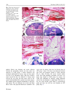
Fig. 2 Histology observation of
laminectomy site in FAM (a),
CAM (b), non-treatment (c),
and AFF groups (d) by H&E
staining at 1 week
postoperatively (9100; CAM
cross-linked amniotic
membrane, FAM freeze–dried
amniotic membrane, EH
epidural hemorrhage, DM dura
mater, NC neural cord, FT fat
tissue)
epidural fibrosis was abundant and severe adhesion nerve roots. In contrast, there was less fibrotic tissue in
between the surrounding muscles and dura mater was AFF group. A layer of fat tissue was interposed between
observed. In CAM group, a fibrous membrane was dura mater and paraspinal muscles to reduce adhesion
developed underneath the laminar edge and dura mater (Fig. 3). After 12 weeks, the results showed similar
was free of scar attachment. Only a thin layer of fibrous trend. The FAM degraded completely and dense scar
tissue around the dura was observed. A distinct layer tissue thus formed in FAM group. In contrast, the
between the scar tissue and dura mater could be dis- CAM still maintained its integrity, which efficiently
cerned which facilitated the exposure in revision surgery. reduced the scar formation in spinal canal. With time on,
The epidural fibrosis appeared prominently in non-treat- the scar tissue became much denser in non-treatment
ment group. The scar adhesion was severer and dense group and there was still less scar formation in AFF
fibrous tissue with populated fibroblasts extended to the group (Fig. 4).
123