Page 285 - Medicine and Surgery
P. 285
P1: KPE
         BLUK007-06  BLUK007-Kendall  May 25, 2005  18:6  Char Count= 0


                                                                         Chapter 6: Genitourinary oncology 281


                   iv Chemotherapy is not as effective and is used mainly  Aetiology
                     for non-responsive disease.                Maldescent of the testis has a 10–15-fold risk. Ten per
                    Throughout treatment a multidisciplinary approach  cent of all testicular tumours develop in testes which

                    is needed with regard to palliation of symptoms. Bis-  are or were cryptorchid, some contra-laterally. A family
                    phosphonates are used for bone pain and to prevent  history is also a known risk factor as is infertility.
                    fractures. Localised radiation is used for bone pain
                    and recently bone-targeting radioisotopes have been
                                                                Pathophysiology
                    developed for those with multiple metastases.
                                                                It is currently thought that the precursor of most germ
                                                                cell tumours is intratubular germ cell neoplasia (some-
                  Prognosis                                     times called testicular carcinoma in situ), where the sem-
                  Depends on grade, volume of primary and TNM stag-  iniferous tubules have atypical germ cells.
                  ing; 50% present with incurable disease. If confined to  It appears that these atypical cells are formed early
                  prostate: 80% have 5-year survival and 60% have 10-  in gestation and may be influenced by events in utero.
                  year survival. If metastases are present: 20% have 5-year  They then lie dormant, until puberty, when they spread
                  survival and 10% have 10-year survival.       non-invasively. In some individuals, they become ma-
                                                                lignant and either develop along the seminomatous or
                                                                teratomatous line.
                  Introduction to testicular tumours

                  Definition
                                                                Classification
                  Tumours of the testis may be classified broadly into those
                                                                The main components of the testis are the germ cells
                  arising from the germ-cell line and those arising from
                                                                (spermatogonia), the sex cords or seminiferous tubules
                  non-germ cells.
                                                                (Sertoli cells) and stroma (Leydig cells). Germ cell tu-
                                                                mours are the most common (90–95%) testicular tu-
                  Incidence                                     mours. Germ cells are multipotent, i.e. can form many
                  Relatively uncommon (∼3–6/100,000 per annum), but  tissue types, as normally they are involved in reproduc-
                  stillthemostcommonsolidorgantumourinyoungmen  tionandmayformbothembryonicandextra-embryonic
                  and increasing in recent years.               tissue. Therefore many different cell types may coexist
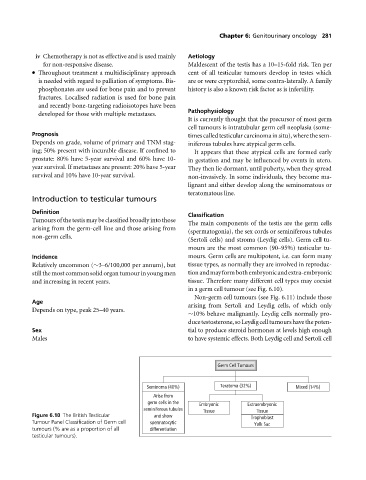
                                                                in a germ cell tumour (see Fig. 6.10).
                                                                 Non-germ cell tumours (see Fig. 6.11) include those
                  Age
                                                                arising from Sertoli and Leydig cells, of which only
                  Depends on type, peak 25–40 years.
                                                                ∼10% behave malignantly. Leydig cells normally pro-
                                                                ducetestosterone,soLeydigcelltumourshavethepoten-
                  Sex                                           tial to produce steroid hormones at levels high enough
                  Males                                         to have systemic effects. Both Leydig cell and Sertoli cell


                                                                        Germ Cell Tumours


                                                    Seminoma (40%)       Teratoma (32%)        Mixed (14%)
                                                     Arise from
                                                    germ cells in the  Embryonic  Extraembryonic
                                                   seminiferous tubules  Tissue    Tissue
                  Figure 6.10 The British Testicular  and show                    Trophoblast
                  Tumour Panel Classification of Germ cell  spermatocytic           Yolk Sac
                  tumours (% are as a proportion of all  differentiation
                  testicular tumours).

