
Page 287 - Medicine and Surgery
P. 287
P1: KPE
BLUK007-06 BLUK007-Kendall May 25, 2005 18:6 Char Count= 0
Chapter 6: Genitourinary oncology 283
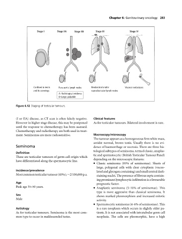
Stage I Stage IIA Stage IIB Stage III Stage IV
Confined to testis Para-aortic lymph nodes Mediastinal and/or Visceral metastases
and its coverings supraclavicular lymph nodes
A–Radiological evidence
B–Large, palpable
Figure 6.12 Staging of testicular tumours.
(I or IIA) disease, as CT scan is often falsely negative. Clinical features
Howeverin higher stage disease, this may be postponed As for testicular tumours. Bilateral involvement is rare.
until the response to chemotherapy has been assessed.
Chemotherapy and radiotherapy are both used in treat-
ment. Seminomas are more radiosensitive. Macroscopy/microscopy
The tumour appears as a homogeneous firm white mass,
amidst normal, brown testis. Usually there is no evi-
Seminoma dence of haemorrhage or necrosis. There are three his-
tological subtypes of seminoma, termed classic, anaplas-
Definition
tic and spermatocytic (British Testicular Tumour Panel)
These are testicular tumours of germ-cell origin which
depending on the microscopic features:
have differentiated along the spermatocytic line.
Classic seminoma (85% of seminomas). Sheets of
large, polygonal cells with clear cytoplasm (vacuo-
Incidence/prevalence latedandglycogencontaining)andsmallcentraldark-
Mostcommontesticulartumour(40%);∼2/100,000p.a. staining nuclei. The presence of fibrous septa contain-
ing prominent lymphocytic infiltration is a favourable
Age
prognostic factor.
Peak age 35–50 years. Anaplastic seminoma (5–10% of seminomas). This
type is more aggressive than classical seminoma. It
Sex
shows marked pleomorphism and increased mitotic
Male
activity.
Spermatocytic seminoma (4–6% of seminomas). This
Aetiology is a rare neoplasm which occurs in slightly older pa-
As for testicular tumours. Seminoma is the most com- tients. It is not associated with intratubular germ cell
mon type to occur in maldescended testes. neoplasia. The cells are pleomorphic, have a high