
Page 212 - Zoo Animal Learning and Training
P. 212
Chapter 25: Vertebral Fracture and Luxation Repair 217
A B
C
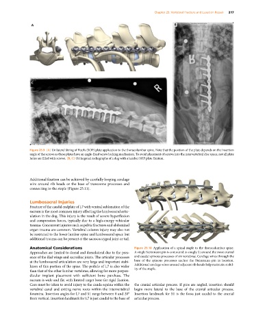
Figure 25.9 (A) Unilateral String of Pearls (SOP) plate application to the thoracolumbar spine. Note that the position of the plate depends on the insertion
angle of the screws as these plates have an angle‐fixed screw locking mechanism. To avoid placement of screws into the intervertebral disc space, not all plate
holes are filled with screws. (B, C) Orthogonal radiographs of a dog with a lumbar SOP plate fixation.
Additional fixation can be achieved by carefully looping cerclage
wire around rib heads or the base of transverse processes and
connecting to the staple (Figure 25.11).
Lumbosacral Injuries
Fracture of the caudal endplate of L7 with ventral subluxation of the
sacrum is the most common injury affecting the lumbosacral artic
ulation in the dog. This injury is the result of severe hyperflexion
and compression forces, typically due to a high‐energy vehicular
trauma. Concurrent injuries such as pelvic fractures and abdominal
organ trauma are common. Vertebral column injury may also not
be restricted to the lower lumbar spine and lumbosacral space but
additional trauma can be present at the sacrococcygeal joint or tail.
Anatomical Considerations Figure 25.10 Application of a spinal staple to the thoracolumbar spine.
Approaches are limited to dorsal and dorsolateral due to the pres A single Steinmann pin is contoured to snugly fit around the most cranial
ence of the ilial wings and sacroiliac joints. The articular processes and caudal spinous processes of six vertebrae. Cerclage wires through the
at the lumbosacral articulation are very large and important stabi base of the spinous processes anchor the Steinmann pin in location.
lizers of this portion of the spine. The pedicle of L7 is also wider Additional cerclage wires around adjacent rib heads help maintain stabil
than that of the other lumbar vertebrae, allowing for more perpen ity of the staple.
dicular implant placement with sufficient bone purchase. The
sacrum is wide and flat with limited target bone for rigid fixation.
Care must be taken to avoid injury to the cauda equina within the the cranial articular process. If pins are angled, insertion should
vertebral canal and exiting nerve roots within the intervertebral begin more lateral to the base of the cranial articular process.
foramina. Insertion angles for L7 and S1 range between 0 and 20° Insertion landmark for S1 is the fossa just caudal to the cranial
from vertical. Insertion landmark for L7 is just caudal to the base of articular process.