
Page 214 - Zoo Animal Learning and Training
P. 214
Chapter 25: Vertebral Fracture and Luxation Repair 219
can be achieved, temporary or permanent transarticular pins or
screws can be placed to help maintain position and free up space
for definitive stabilization. If reduction cannot be achieved and
subluxation causes compression of the cauda equina by the sacral
lamina, a partial dorsal laminectomy can be carefully performed to
remove the compressing bone.
Another anchor point for reduction forceps are the ilial wings,
which can provide a larger area for instruments to attach to and
manipulate the sacrum.
Pins or Screws and PMMA
Insertion angle and landmarks are reviewed and adjusted based on
patient‐specific anatomy. Positive‐profile pins are preferred over
cortical screws due to their superior stiffness. Most medium to large
dogs will accept ⅛ inch pins or 3.5 mm cortical screws. Principles of
application are the same as for thoracolumbar implants. L7 and S1
pins can be slightly angled in a cranial and caudal direction to bring
the protruding pin portions into closer proximity for PMMA appli
cation (Figure 25.12). If needed, pins can be bent carefully toward
the L7–S1 disc space to improve pin incorporation into the cement;
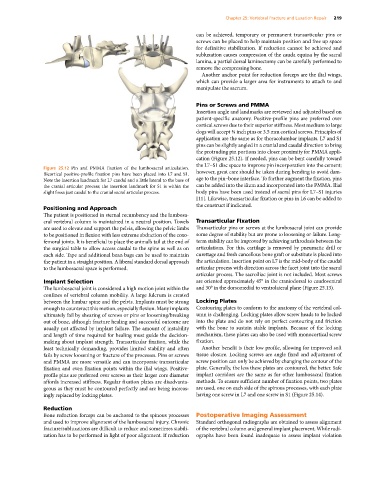
Figure 25.12 Pin and PMMA fixation of the lumbosacral articulation.
Bicortical positive‐profile fixation pins have been placed into L7 and S1. however, great care should be taken during bending to avoid dam
Note the insertion landmark for L7 caudal and a little lateral to the base of age to the pin–bone interface. To further augment the fixation, pins
the cranial articular process; the insertion landmark for S1 is within the can be added into the ilium and incorporated into the PMMA. Ilial
slight fossa just caudal to the cranial sacral articular process. body pins have been used instead of sacral pins for L7–S1 injuries
[11]. Likewise, transarticular fixation or pins in L6 can be added to
the construct if indicated.
Positioning and Approach
The patient is positioned in sternal recumbency and the lumbosa
cral vertebral column is maintained in a neutral position. Towels Transarticular Fixation
are used to elevate and support the pelvis, allowing the pelvic limbs Transarticular pins or screws at the lumbosacral joint can provide
to be positioned in flexion with less extreme abduction of the coxo some degree of stability but are prone to loosening or failure. Long‐
femoral joints. It is beneficial to place the animal’s tail at the end of term stability can be improved by achieving arthrodesis between the
the surgical table to allow access caudal to the spine as well as on articulations. For this, cartilage is removed by pneumatic drill or
each side. Tape and additional bean bags can be used to maintain curettage and fresh cancellous bone graft or substitute is placed into
the patient in a straight position. A liberal standard dorsal approach the articulation. Insertion point on L7 is the mid‐body of the caudal
to the lumbosacral space is performed. articular process with direction across the facet joint into the sacral
articular process. The sacroiliac joint is not included. Most screws
Implant Selection are oriented approximately 45° in the craniodorsal to caudoventral
The lumbosacral joint is considered a high motion joint within the and 30° in the dorsomedial to ventrolateral plane (Figure 25.13).
confines of vertebral column mobility. A large fulcrum is created
between the lumbar spine and the pelvis. Implants must be strong Locking Plates
enough to counteract this motion, especially flexion. Many implants Contouring plates to conform to the anatomy of the vertebral col
ultimately fail by shearing of screws or pins or loosening/breaking umn is challenging. Locking plates allow screw heads to be locked
out of bone, although fracture healing and successful outcome are into the plate and do not rely on perfect contouring and friction
usually not affected by implant failure. The amount of instability with the bone to sustain stable implants. Because of the locking
and length of time required for healing must guide the decision‐ mechanism, these plates can also be used with monocortical screw
making about implant strength. Transarticular fixation, while the fixation.
least technically demanding, provides limited stability and often Another benefit is their low profile, allowing for improved soft
fails by screw loosening or fracture of the processes. Pins or screws tissue closure. Locking screws are angle fixed and adjustment of
and PMMA are more versatile and can incorporate transarticular screw position can only be achieved by changing the contour of the
fixation and even fixation points within the ilial wings. Positive‐ plate. Generally, the less these plates are contoured, the better. Safe
profile pins are preferred over screws as their larger core diameter implant corridors are the same as for other lumbosacral fixation
affords increased stiffness. Regular fixation plates are disadvanta methods. To ensure sufficient number of fixation points, two plates
geous as they must be contoured perfectly and are being increas are used, one on each side of the spinous processes, with each plate
ingly replaced by locking plates. having one screw in L7 and one screw in S1 (Figure 25.14).
Reduction
Bone reduction forceps can be anchored to the spinous processes Postoperative Imaging Assessment
and used to improve alignment of the lumbosacral injury. Chronic Standard orthogonal radiographs are obtained to assess alignment
fracture/subluxations are difficult to reduce and sometimes stabili of the vertebral column and general implant placement. While radi
zation has to be performed in light of poor alignment. If reduction ographs have been found inadequate to assess implant violation