
Page 389 - Adams and Stashak's Lameness in Horses, 7th Edition
P. 389
Diagnostic Imaging 355
VetBooks.ir
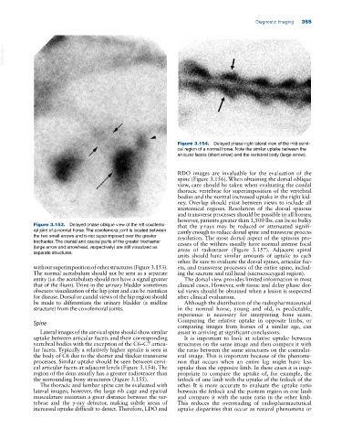
Figure 3.154. Delayed phase right lateral view of the mid‐cervi
cal region of a normal horse. Note the similar uptake between the
articular facets (short arrow) and the vertebral body (large arrow).
RDO images are invaluable for the evaluation of the
spine (Figure 3.156). When obtaining the dorsal oblique
view, care should be taken when evaluating the caudal
thoracic vertebrae for superimposition of the vertebral
bodies and the normal increased uptake in the right kid
ney. Overlap should exist between views to include all
anatomical regions. Resolution of the dorsal spinous
and transverse processes should be possible in all horses;
however, patients greater than 1,500 lbs. can be so bulky
Figure 3.153. Delayed phase oblique view of the left coxofemo that the γ‐rays may be reduced or attenuated signifi
ral joint of a normal horse. The coxofemoral joint is located between cantly enough to reduce dorsal spine and transverse process
the two small arrows and is not superimposed over the greater resolution. The most dorsal aspect of the spinous pro
trochanter. The cranial and caudal parts of the greater trochanter cesses of the withers usually have normal intense focal
(large arrow and arrowhead, respectively) are still visualized as areas of radiotracer (Figure 3.157). Adjacent spinal
separate structures.
units should have similar amounts of uptake to each
other. Be sure to evaluate the dorsal spines, articular fac
without superimposition of other structures (Figure 3.153). ets, and transverse processes of the entire spine, includ
The normal acetabulum should not be seen as a separate ing the sacrum and tail head (sacrococcygeal region).
entity (i.e. the acetabulum should not have a signal greater The dorsal view provides limited information in most
that of the ilium). Urine in the urinary bladder sometimes clinical cases. However, soft tissue and delay phase dor
obscures visualization of the hip joint and can be mistaken sal views should be obtained when a lesion is suspected
for disease. Dorsal or caudal views of the hip region should after clinical evaluation.
be made to differentiate the urinary bladder (a midline Although the distribution of the radiopharmaceutical
structure) from the coxofemoral joints. in the normal horse, young and old, is predictable,
experience is necessary for interpreting bone scans.
Spine Comparing the relative uptake in opposite limbs, or
comparing images from horses of a similar age, can
Lateral images of the cervical spine should show similar assist in arriving at significant conclusions.
uptake between articular facets and their corresponding It is important to look at relative uptake between
vertebral bodies with the exception of the C6–C7 articu structures on the same image and then compare it with
lar facets. Typically a relatively higher uptake is seen in the ratio between the same structures on the contralat
the body of C6 due to the shorter and thicker transverse eral image. This is important because of the phenome
processes. Similar uptake should be seen between cervi non that occurs when an entire leg might have less
cal articular facets at adjacent levels (Figure 3.154). The uptake than the opposite limb. In these cases it is inap
region of the dens usually has a greater radiotracer than propriate to compare the uptake of, for example, the
the surrounding bony structures (Figure 3.155). fetlock of one limb with the uptake of the fetlock of the
The thoracic and lumbar spine can be evaluated with other. It is more accurate to evaluate the uptake ratio
lateral images; however, the large rib cage and epaxial between the fetlock and the pastern region in one limb
musculature maintain a great distance between the ver and compare it with the same ratio in the other limb.
tebrae and the γ‐ray detector, making subtle areas of This reduces the overreading of radiopharmaceutical
increased uptake difficult to detect. Therefore, LDO and uptake disparities that occur as natural phenomena or