
Page 393 - Adams and Stashak's Lameness in Horses, 7th Edition
P. 393
Diagnostic Imaging 359
VetBooks.ir
A B
Figure 3.162. (A) Delayed phase lateral view of the left tarsus the well‐defined radiolucent fracture line extending from the
of a horse with focal and intense abnormal radiotracer in the proximal to the distal articular surfaces of the third tarsal bone,
region of the distal tarsus corresponding to a third tarsal bone consistent with a slab fracture (arrow). Source: Courtesy of
fracture. (B) Lateromedial radiograph of the same tarsus showing Dr. Erik Bergman.
traumatic fractures less than 24 hours’ duration fail to
show increased tracer uptake when compared with
adjacent bone. Figure 3.163 is a delayed phase image
of a comminuted middle phalangeal fracture 48 hours’
post‐injury. Although mild increased uptake is seen, the
fracture is best diagnosed due to anatomic abnormality
and not its physiologic peculiarity. Compare this
with Figure 3.161, a chronic proximal phalangeal frac
ture with intense uptake but minimal anatomic dis
placement. Fracture uptake in humans is expected at
about 24 hours’ post‐injury (although it takes longer
in older patients) and is expected to last for 6–12
months or longer in older patients. The uptake by a
88
fracture should decrease over time as fracture healing
occurs.
Multifocal areas of abnormal radiotracer have been
described with different diseases such as enostosis‐like
50
lesions, 7,66 hypertrophic osteopathy, neoplasia, 27,43 and
horses with a bone fragility disorder, a recently reported
2
condition of unknown etiology that affects the axial and
proximal appendicular skeleton. Figure 3.164 shows
intestinal adenocarcinoma metastases to the ribs and
distal left humerus. The same horse also had metastatic
disease to several cervical, thoracic, and lumbar vertebrae,
multiple ribs, and the sternum.
Localized delayed phase uptake of the radiophar
maceutical by soft tissues is not commonly seen and
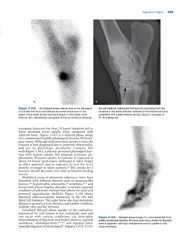
can occur with various conditions, e.g. dystrophic Figure 3.163. Delayed phase image of a comminuted left front
mineralization of ligament and tendon injuries, regional middle phalangeal fracture 48 hours’ post‐injury. Anatomic displace
anesthesia, rhabdomyolysis, 13,41,59 and repeated intra ment is apparent, although radiopharmaceutical uptake is only
1
muscular injection of butorphanol (Figures 3.133, 3.165, mildly increased.
49