
Page 414 - Adams and Stashak's Lameness in Horses, 7th Edition
P. 414
380 Chapter 3
VetBooks.ir
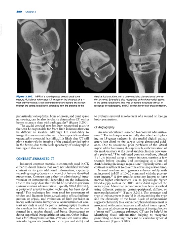
Figure 3.197. MPR of a non‐displaced central tarsal bone distal articular surface, with a dorsomedial to plantarolateral orienta
fracture.Multiplanar reformatted CT images of the left tarsus of a 7‐ tion. (Arrows) Sclerosis is also recognized at the dorsomedial aspect
year‐old Warmblood. A well‐defined radiolucent fracture line is seen of the central tarsal bone. This type of fracture is typically difficult to
through the central tarsal bone, extending from the proximal to the recognize on radiographs, and CT is often key in their characterization.
periarticular osteophytes, bone sclerosis, and joint space to evaluate synovial involvement of a wound or foreign
narrowing, can be also be clearly detected on CT with a body penetration.
better accuracy than with radiographs (Figure 3.200).
18
The caudal cervical area has been recognized as a site CT Angiography
that can be responsible for front limb lameness that can
be difficult to localize. Although CT availability to An arterial catheter is needed for contrast administra
image this area remains limited, a few reports have dem tion. The technique was initially described with plac
19
onstrated its potential feasibility. It is likely that CT will ing an 18‐gauge catheter in the medial digital palmar
play a major role in imaging of the caudal cervical spine artery just distal to the carpus using ultrasound guid
in the future, due to the lack specificity of radiographic ance. Due to occasional poor perfusion of the lateral
findings of this area. aspect of the foot using this approach, catheterization of
the median artery at the distal antebrachium is now usu
ally preferred. The iodinated contrast medium, diluted
9
CONTRAST‐ENHANCED CT 1 : 1, is injected using a power injector, starting a few
seconds before imaging and continuing at a rate of
Iodinated contrast material is commonly used in CT, 2 mL/s during the image acquisition. Typically 60 mL of
19
either to detect lesions that were not identified without contrast is sufficient to cover the entire acquisition time.
contrast or to gain additional information, especially Normal tendons and ligaments enhance mildly with
regarding staging (acute vs. chronic) of lesions identified an increased in HU of 10–20 compared with the precon
precontrast. Contrast can either be administered intra trast images. A few specific areas are known to have
19
vascular or intrasynovial depending on the indication. normal higher enhancement due to physiologic higher
Due to the large dose that would be needed to perform blood supply, such as the DDFT at the distal aspect of the
systemic contrast administration (typically 500–1,000 mL), metacarpus. Abnormal enhancement has been described
a peripheral arterial injection technique has been devel using different patterns: central–peripheral, diffuse, or
oped. This technique has been used for assessment of neovascularization 9,24 (Figure 3.201). The presence and
4
tendon and ligament lesions, evaluation of local inflam type of enhancement is useful in characterizing the type
mation or sepsis, and evaluation of limb perfusion in and the chronicity of the lesion. Lack of enhancement
horses with laminitis. Intrasynovial administration of con suggests chronicity to a lesion. Peripheral enhancement is
trast not only is used for joints (arthrography) to assess associated with central necrosis and lack of tissue repair. 9
the cartilage for defects or thinning but also can be per In cases of abscesses or penetration of foreign bodies
formed in a tendon sheath and bursa (bursogram) to (“street nail”), contrast‐enhanced CT can be helpful in
detect superficial irregularities of tendons. Other indica identifying focal inflammation helping to recognize
tions for intrasynovial administration is to assess intra‐ penetrating or draining tracts and to assess for synovial
articular ligaments (mostly in the carpus and stifle) and involvement (Figure 3.202).