
Page 417 - Adams and Stashak's Lameness in Horses, 7th Edition
P. 417
Diagnostic Imaging 383
VetBooks.ir
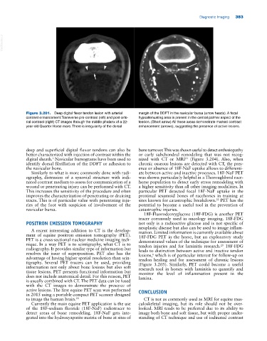
Figure 3.201. Deep digital flexor tendon lesion with arterial margin of the DDFT in the navicular bursa (arrow heads). A focal
contrast enhancement.Transverse pre‐contrast (left) and post‐arte hypoattenuating area is present in the central palmar aspect of the
rial contrast (right) CT images through the middle phalanx of a 22‐ tendon. (Short arrow) All these areas demonstrate marked contrast
year‐old Quarter Horse mare. There is irregularity of the dorsal enhancement (arrows), suggesting the presence of active lesions.
deep and superficial digital flexor tendons can also be bone turnover. This was shown useful to detect enthesiopathy
better characterized with injection of contrast within the or early subchondral remodeling that was not recog
21
1
digital sheath. Navicular bursograms have been used to nized with CT or MRI (Figure 3.204). Also, when
identify dorsal fibrillation of the DDFT or adhesion to chronic osseous lesions are detected with CT, the pres
the navicular bone. ence or absence of 18F‐NaF uptake allows to differenti
Similarly to what is more commonly done with radi ate between active and inactive processes. 18F‐NaF PET
ography, distension of a synovial structure with iodi was shown particularly helpful in a Thoroughbred race
nated contrast medium to assess for communication of a horse population to detect early stress remodeling with
wound or penetrating injury can be performed with CT. a higher sensitivity than all other imaging modalities. In
This increases the sensitivity of the procedure and often particular PET detected focal 18F‐NaF uptake in the
improves the characterization of penetrating or draining proximal sesamoid bones of racehorses in training at
tracts. This is of particular value with penetrating inju sites known for catastrophic breakdown. PET has the
22
ries of the foot with suspicion of involvement of the potential to become a useful tool in the prevention of
navicular bursa. catastrophic injuries.
18F‐Fluorodeoxyglucose (18F‐FDG) is another PET
tracer commonly used in oncology imaging. 18F‐FDG
POSITRON EMISSION TOMOGRAPHY not only is a radioactive glucose and is not specific of
neoplastic disease but also can be used to image inflam
A recent interesting addition to CT is the develop mation. Limited information is currently available about
ment of equine positron emission tomography (PET). 18F‐FDG PET in the horse, but an exploratory study
PET is a cross‐sectional nuclear medicine imaging tech demonstrated values of the technique for assessment of
nique. In a way PET is to scintigraphy, what CT is to tendon injuries and for laminitis research. 18F‐FDG
20
radiography. It provides similar type of information but leads to distinction between active and inactive tendon
resolves the issue of superposition. PET also has the lesions, which is of particular interest for follow‐up on
6
advantage of having higher spatial resolution than scin tendon healing and for assessment of chronic lesions
tigraphy. Several PET tracers can be used, providing (Figure 3.205). Similarly, PET could become a useful
information not only about bone lesions but also soft research tool in horses with laminitis to quantify and
tissue lesions. PET presents functional information but monitor the level of inflammation present in the
does not include anatomical detail. For this reason, PET lamina.
is usually combined with CT. The PET data can be fused
with the CT images to demonstrate the presence of
active lesions. The first equine PET scan was performed CONCLUSION
in 2015 using a portable compact PET scanner designed
to image the human brain. 20 CT is not as commonly used as MRI for equine mus
Currently the main equine PET application is the use culoskeletal imaging, but its role should not be over
of the 18F‐sodium fluoride (18F‐NaF) radiotracer to looked. MRI tends to be preferred due to its ability to
detect areas of bone remodeling. 18F‐NaF gets inte image both bone and soft tissue, but with proper under
grated into the hydroxyapatite matrix of bone at sites of standing of CT technique and use of iodinated contrast