
Page 422 - Adams and Stashak's Lameness in Horses, 7th Edition
P. 422
388 Chapter 3
VetBooks.ir
Figure 3.206. A Siemens Magnetom Skyra 3T high‐field with the lame limb lowermost. The region of interest in the limb, in
magnet (Siemens, Malvern, PA) used for MRI of horses under this case the foot, is positioned in the isocenter of the magnet.
general anesthesia. The horse is positioned in lateral recumbency
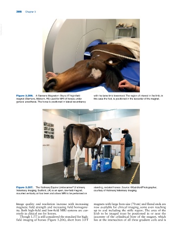
Figure 3.207. The Hallmarq Equine Limbscanner (Hallmarq standing, sedated horses. Source: ©GuildfordPhotographer,
®
Veterinary Imaging, Guilford, UK) is an open, low‐field magnet, courtesy of Hallmarq Veterinary Imaging.
mounted vertically at floor level and allows MRI to be performed on
Image quality and resolution increase with increasing magnets with large bore size (70 cm) and flared ends are
magnetic field strength and increasing field homogene now available for clinical imaging, some even reaching
ity. Both high‐field and low‐field MRI systems are cur up to and including the stifle region. The area of the
rently in clinical use for horses. limb to be imaged must be positioned in or near the
Though 1.5 T is still considered the standard for high‐ isocenter of the cylindrical bore of the magnet, which
field imaging of horses (Figure 3.206), short bore 3.0 T lies at the intersection of all three gradient coils and is