
Page 426 - Adams and Stashak's Lameness in Horses, 7th Edition
P. 426
392 Chapter 3
VetBooks.ir
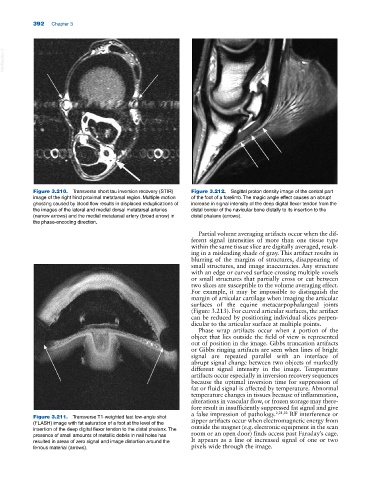
Figure 3.210. Transverse short tau inversion recovery (STIR) Figure 3.212. Sagittal proton density image of the central part
image of the right hind proximal metatarsal region. Multiple motion of the foot of a forelimb. The magic angle effect causes an abrupt
ghosting caused by blood flow results in displaced reduplications of increase in signal intensity of the deep digital flexor tendon from the
the images of the lateral and medial dorsal metatarsal arteries distal border of the navicular bone distally to its insertion to the
(narrow arrows) and the medial metatarsal artery (broad arrow) in distal phalanx (arrows).
the phase‐encoding direction.
Partial volume averaging artifacts occur when the dif
ferent signal intensities of more than one tissue type
within the same tissue slice are digitally averaged, result
ing in a misleading shade of gray. This artifact results in
blurring of the margins of structures, disappearing of
small structures, and image inaccuracies. Any structure
with an edge or curved surface crossing multiple voxels
or small structures that partially cross or cut between
two slices are susceptible to the volume averaging effect.
For example, it may be impossible to distinguish the
margin of articular cartilage when imaging the articular
surfaces of the equine metacarpophalangeal joints
(Figure 3.213). For curved articular surfaces, the artifact
can be reduced by positioning individual slices perpen
dicular to the articular surface at multiple points.
Phase wrap artifacts occur when a portion of the
object that lies outside the field of view is represented
out of position in the image. Gibbs truncation artifacts
or Gibbs ringing artifacts are seen when lines of bright
signal are repeated parallel with an interface of
abrupt signal change between two objects of markedly
different signal intensity in the image. Temperature
artifacts occur especially in inversion recovery sequences
because the optimal inversion time for suppression of
fat or fluid signal is affected by temperature. Abnormal
temperature changes in tissues because of inflammation,
alterations in vascular flow, or frozen storage may there
fore result in insufficiently suppressed fat signal and give
a false impression of pathology. 2,21,22 RF interference or
Figure 3.211. Transverse T1‐weighted fast low‐angle shot zipper artifacts occur when electromagnetic energy from
(FLASH) image with fat saturation of a foot at the level of the outside the magnet (e.g. electronic equipment in the scan
insertion of the deep digital flexor tendon to the distal phalanx. The
presence of small amounts of metallic debris in nail holes has room or an open door) finds access past Faraday’s cage.
resulted in areas of zero signal and image distortion around the It appears as a line of increased signal of one or two
ferrous material (arrows). pixels wide through the image.