
Page 147 - Basic Monitoring in Canine and Feline Emergency Patients
P. 147
(A) (C)
VetBooks.ir R R PPL R TI R PPL
A A A A A
(E)
PPL
R NOD R
(B) (D) A A
PPL Sh
R R PPL
R Shrd R
B
A A A A
B
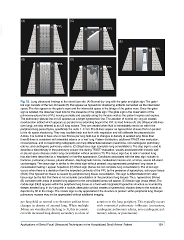
Fig. 7.6. Lung ultrasound findings at the chest tube site. (A) Normal dry lung with the gator and glide sign: The gator/
bat sign consists of the two rib heads (R) that appear as hypoechoic shadowing artifacts connected via the intercostal
space. The ribs appear as the gator’s eyes and the intercostal space is the bridge of the gators nose. Once the gator
sign is isolated, the observer must look for the presence of the ‘glide sign.’ The glide sign is the observation of the
pulmonary–pleural line (PPL) moving cranially and caudally along the thoracic wall as the patient inspires and expires.
The pulmonary–pleural line on US appears as a bright hyperechoic line. The aeration of normal dry lung air creates
reverberation artifact which appears as parallel lines extending beyond the PPL termed A-lines (A). (B) Ultrasound B-lines
(wet lung); are also referred to as US lung rockets. They are created when fluid is immediately next to air within the
peripheral lung parenchyma, specifically the outer 1–3 mm. The B-lines appear as hyperechoic streaks that run parallel
to the rib space shadowing. They may oscillate back and forth with respiration and will obliterate the perpendicular
A-lines. It is normal to have one or two B-lines per lung field due to changes in density of aerated lung. More than
three B-lines is consistent with interstitial edema or a ‘wet’ lung. Pattern distribution, additional TFAST view evaluation,
clinical picture, and corresponding radiographs can help differentiate between pneumonia, non-cardiogenic pulmonary
edema, and cardiogenic pulmonary edema. (C) Step/tissue sign (complete lung consolidation). The step sign is used to
describe a discontinuity in the pulmonary–pleural line during TFAST evaluation, usually associated with thoracic wall
3
or pleural space disease and/or lung consolidation without aeration (TI). The tissue sign due to lack of aerated lung
has also been described as a ‘hepatized’ or liver-like appearance. Conditions associated with the step sign include rib
fractures, pulmonary masses, pleural effusion, diaphragmatic hernia, mediastinal masses and, at times, severe left-sided
cardiomegaly. The tissue sign is similar to the shred sign without aerated lung appreciated; peripheral lung tissue is
consolidated making it appear hepatized. (D) Shred sign (dense but not complete lung consolidation). The shred sign
occurs when there is a deviation from the expected continuity of the PPL by the presence of hypoechoic, echo-poor tissue
(Shrd). This hypoechoic tissue is caused by peripheral lung tissue consolidation. This sign is differentiated from step/
tissue sign by the fact that there is not complete consolidation of the peripheral lung tissues. Thus, hyperechoic B-lines
(B) consistent with areas of aerated lung adjacent to the consolidated areas still appear. (E) Nodule sign (mass effect). The
nodule sign (Nod) appears within the peripheral lung tissue as a focal well marginated hypoechoic structure surrounded by
deeper aerated lung. In dry lung with a nodule, attenuation artifact creates a hyperechoic shadow deep to the nodule as
depicted by Sh in the image. The nodule sign is only appreciated if the structure is present within peripheral lung. Deeper
pulmonary masses may not be appreciated without additional imaging.
per lung field as normal reverberation artifact from aeration in the lung periphery. This typically occurs
changes in density of aerated lung. When multiple with interstitial pulmonary infiltrates (contusions,
B-lines are visualized in the lung (> 3), this is consist- cardiogenic pulmonary edema, non-cardiogenic pul-
ent with increased lung density secondary to a loss of monary edema, or pneumonia).
Applications of Serial Focal Ultrasound Techniques in the Hospitalized Small Animal Patient 139