
Page 149 - Basic Monitoring in Canine and Feline Emergency Patients
P. 149
(A) (B) FR 27
AO% 100
LOGIQ 0 LOGIQ RV 0 Frq 8.0
B
S8
VetBooks.ir RV RA Ao PA 2 Gn D/0
S8
60
3/2
S/A
Map
D
8.0
DR
90
LV 2
4
LA
LA
6
4
8
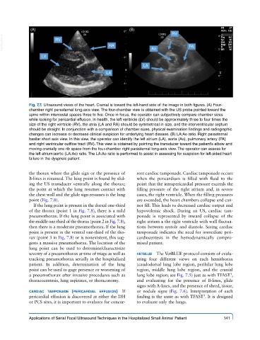
Fig. 7.7. Ultrasound views of the heart. Cranial is toward the left-hand side of the image in both figures. (A) Four-
chamber right parasternal long-axis view. The four-chamber view is obtained with the US probe pointed toward the
spine within intercostal spaces three to five. Once in focus, the operator can subjectively compare chamber sizes
while looking for pericardial effusion. In health, the left ventricle (LV) should be approximately three to four times the
size of the right ventricle (RV), the atria (LA and RA) should be symmetrical in size, and the interventricular septum
should be straight. In conjunction with a comparison of chamber sizes, physical examination findings and radiographic
changes can increase or decrease clinical suspicion for underlying heart disease. (B) LA:Ao ratio. Right parasternal
basilar short axis view. In this view, the operator can identify the left atrium (LA), aorta (Ao), pulmonary artery (PA)
and right ventricular outflow tract (RV). This view is obtained by pointing the transducer toward the patient’s elbow and
moving cranially one rib space from the four-chamber right parasternal long-axis view. The operator can assess for
the left atrium:aortic (LA:Ao) ratio. The LA:Ao ratio is performed to assist in assessing for suspicion for left-sided heart
failure in the dyspneic patient.
the thorax where the glide sign or the presence of rent cardiac tamponade. Cardiac tamponade occurs
B-lines is resumed. The lung point is found by slid- when the pericardium is filled with fluid to the
ing the US transducer ventrally along the thorax; point that the intrapericardial pressure exceeds the
the point at which the lung resumes contact with filling pressure of the right atrium and, in severe
the chest wall and the glide sign resumes is the lung cases, the right ventricle. When the filling pressures
point (Fig. 7.8). are exceeded, the heart chambers collapse and can-
If the lung point is present in the dorsal one-third not fill. This leads to decreased cardiac output and
of the thorax (point 1 in Fig. 7.8), there is a mild hypovolemic shock. During an US, cardiac tam-
pneumothorax. If the lung point is associated with ponade is represented by inward collapse of the
the middle one-third of the thorax (point 2 in Fig. 7.8), right atrium ± the right ventricle with wall fluctua-
then there is a moderate pneumothorax. If the lung tions between systole and diastole. Seeing cardiac
point is present in the ventral one-third of the tho- tamponade indicates the need for immediate peri-
rax (point 3 in Fig. 7.8) or is nonexistent, this sug- cardiocentesis in the hemodynamically compro-
gests a massive pneumothorax. The location of the mised patient.
lung point can be used to determine/characterize
severity of a pneumothorax at time of triage as well as vetblue The VetBLUE protocol consists of evalu-
tracking pneumothorax serially in the hospitalized ating four different views on each hemithorax
patient. In addition, determination of the lung (caudodorsal lung lobe region, perihilar lung lobe
point can be used to gage presence or worsening of region, middle lung lobe region, and the cranial
a pneumothorax after invasive procedures such as lung lobe region; see Fig. 7.5) just as with TFAST ,
3
thoracocentesis, lung aspirates, or thoracotomy. and evaluating for the presence of B-lines, glide
signs with A-lines, and the presence of shred, tissue,
cardiac tamponade (pericardial effusion) If or nodule signs (Fig. 7.6). Interpretation of each
3
pericardial effusion is discovered at either the DH finding is the same as with TFAST . It is designed
or PCS sites, it is important to evaluate for concur- to evaluate only the lungs.
Applications of Serial Focal Ultrasound Techniques in the Hospitalized Small Animal Patient 141