
Page 154 - Basic Monitoring in Canine and Feline Emergency Patients
P. 154
3
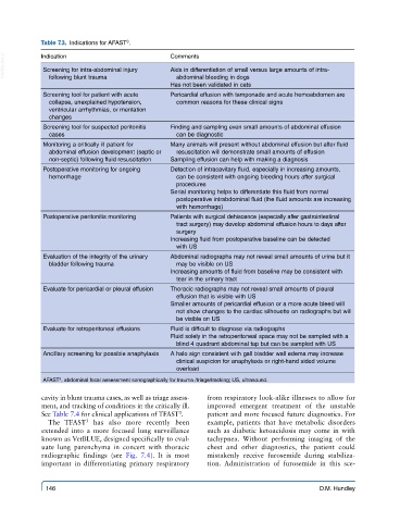
Table 7.3. Indications for AFAST .
VetBooks.ir Indication Comments
Aids in differentiation of small versus large amounts of intra-
Screening for intra-abdominal injury
following blunt trauma
abdominal bleeding in dogs
Has not been validated in cats
Screening tool for patient with acute Pericardial effusion with tamponade and acute hemoabdomen are
collapse, unexplained hypotension, common reasons for these clinical signs
ventricular arrhythmias, or mentation
changes
Screening tool for suspected peritonitis Finding and sampling even small amounts of abdominal effusion
cases can be diagnostic
Monitoring a critically ill patient for Many animals will present without abdominal effusion but after fluid
abdominal effusion development (septic or resuscitation will demonstrate small amounts of effusion
non-septic) following fluid resuscitation Sampling effusion can help with making a diagnosis
Postoperative monitoring for ongoing Detection of intracavitary fluid, especially in increasing amounts,
hemorrhage can be consistent with ongoing bleeding hours after surgical
procedures
Serial monitoring helps to differentiate this fluid from normal
postoperative intrabdominal fluid (the fluid amounts are increasing
with hemorrhage)
Postoperative peritonitis monitoring Patients with surgical dehiscence (especially after gastrointestinal
tract surgery) may develop abdominal effusion hours to days after
surgery
Increasing fluid from postoperative baseline can be detected
with US
Evaluation of the integrity of the urinary Abdominal radiographs may not reveal small amounts of urine but it
bladder following trauma may be visible on US
Increasing amounts of fluid from baseline may be consistent with
tear in the urinary tract
Evaluate for pericardial or pleural effusion Thoracic radiographs may not reveal small amounts of pleural
effusion that is visible with US
Smaller amounts of pericardial effusion or a more acute bleed will
not show changes to the cardiac silhouette on radiographs but will
be visible on US
Evaluate for retroperitoneal effusions Fluid is difficult to diagnose via radiographs
Fluid solely in the retroperitoneal space may not be sampled with a
blind 4 quadrant abdominal tap but can be sampled with US
Ancillary screening for possible anaphylaxis A halo sign consistent with gall bladder wall edema may increase
clinical suspicion for anaphylaxis or right-hand sided volume
overload
AFAST , abdominal focal assessment sonographically for trauma /triage/tracking; US, ultrasound.
3
cavity in blunt trauma cases, as well as triage assess- from respiratory look-alike illnesses to allow for
ment, and tracking of conditions in the critically ill. improved emergent treatment of the unstable
3
See Table 7.4 for clinical applications of TFAST . patient and more focused future diagnostics. For
The TFAST has also more recently been example, patients that have metabolic disorders
3
extended into a more focused lung surveillance such as diabetic ketoacidosis may come in with
known as VetBLUE, designed specifically to eval- tachypnea. Without performing imaging of the
uate lung parenchyma in concert with thoracic chest and other diagnostics, the patient could
radiographic findings (see Fig. 7.4). It is most mistakenly receive furosemide during stabiliza-
important in differentiating primary respiratory tion. Administration of furosemide in this sce-
146 D.M. Hundley