
Page 157 - Basic Monitoring in Canine and Feline Emergency Patients
P. 157
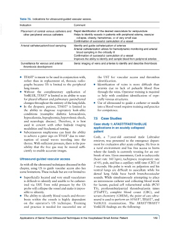
Table 7.6.  Indications for ultrasound-guided vascular access.

  VetBooks.ir  Indication                    Comment
              Placement of central venous catheters and  Rapid identification of the desired vasculature for venipuncture
                                             Helps to identify vessels in patients with peripheral edema, vascular
               other peripheral venous catheters
                                               collapse, obesity, hematomas, or of very small size
                                             Confirmation of successful cannulation of a vessel
              Arterial catheterization/blood sampling  Identify and guide catheterization of arteries
                                             Arterial catheterization allows for hemodynamic monitoring and arterial
                                               blood sampling in the critically ill
                                             Confirmation of successful cannulation of a vessel
                                             Improves the ability to identify and sample blood from peripheral arteries
              Surveillance for venous and arterial   Serial imaging of veins and arteries to identify and describe thrombosis
               thrombosis development


              ● ● TFAST  is meant to be used in conjunction with,   the UST for vascular access and thrombus
                     3
               rather  than in replacement  of, thoracic  radio-  identification.
               graphs because US is limited to the peripheral   ● ● Identification of veins is more difficult than
               lung tissues.                                arteries due to lack of pulsatile blood flow
              ● ● Without the complimentary application  of   through the veins. Operator training is required
               VetBLUE, TFAST  is limited in its ability to scan   to help with confident identification of espe-
                             3
               for pleural effusion and pulmonary parenchymal   cially venous structures.
               changes throughout the entirety of the lung fields.  ● ● Use of ultrasound to guide a catheter or needle
                                         3
              ● ● In the dyspneic patient, TFAST  is limited in   into a blood vessel requires training and practice
               the  ability  to  diagnose  respiratory  look-alike   for competence.
               conditions (examples: diabetic ketoacidosis,
               hypocalcemia, hypoglycemia, hypovolemic shock,   7.5  Case Studies
               and neurologic  disease).  Therefore,  it is best
               used in concert with other bedside imaging   Case study 1: AFAST/TFAST/VetBLUE
               modalities and biochemical testing.       applications in an acutely collapsed
              ● ● Subcutaneous emphysema can limit the ability   patient
               to achieve a gator sign on TFAST  due to inter-  Cash, a 7-year-old castrated male Labrador
                                          3
               ruption of sound waves traveling into the   retriever, was presented to the emergency depart-
               thorax. With sufficient pressure, there is the pos-  ment for evaluation after acute collapse. He lives in
               sibility that the free gas may be moved suffi-  a rural environment and has free access to barns
               ciently to enable accurate images.        where the family is currently treating for an out-
                                                         break of rats. Upon assessment, Cash is tachycardic
             Ultrasound-guided vascular access           (heart rate 160 bpm), tachypneic (respiratory rate
                                                         of 45), pale, and has a capillary refill time (CRT) of
             As with all the ultrasound techniques discussed in this   3 seconds. His pulse is weak on palpation and his
             chapter, using US to guide vascular access also has   ventral lungs are difficult to auscultate while his
             some limitations. These include but are not limited to:
                                                         dorsal lung fields have harsh bronchovesicular
              ● ● Superficially located and very small vasculature   sounds. While simultaneously attempting to place
               is difficult to identify and unable to be catheter-  an intravenous catheter and collecting blood work
               ized via UST. Even mild pressure by the US   for lactate, packed cell volume/total solids (PCV/
               probe will collapse the vessel and make it impos-  TS), prothrombin/partial thromboplastin times
               sible to identify.                        (PT/aPTT), complete blood count  (CBC),  and
              ● ● The ability to identify blood vessels and throm-  serum chemistry (CHEM), the point of care ultra-
                                                                                     3
                                                                                            3
               bosis within the vessels is highly dependent   sound is used to perform an AFAST , TFAST , and
                                                                                         3
               on the operator’s US technique.  Training   VetBLUE  examination. The AFAST /TFAST /
                                                                                                3
               and  practice is needed for successful use of   VetBLUE findings are the following:
             Applications of Serial Focal Ultrasound Techniques in the Hospitalized Small Animal Patient   149