
Page 159 - Basic Monitoring in Canine and Feline Emergency Patients
P. 159
up with his primary care veterinarian in 3 days’
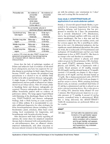
Pericardial site No evidence of No evidence of time and is acting like his normal self.
pericardial
pericardial
VetBooks.ir Symmetrical effusion Case study 3: AFAST/TFAST/VetBLUE
effusion
atrial sizes.
An estimated applications in an acute abdomen patient
LA:Ao ratio is 1 Brindy, a 12-year-old spayed female Sheltie, is pre-
DH No pericardial, pleural or sented to the emergency department for vomiting,
peritoneal effusion appreciated diarrhea, lethargy, and hyporexia that has pro-
gressed to anorexia for 2 days. On presentation,
Caudodorsal lung Glide sign + Glide sign + she is severely dehydrated (~9% dehydration),
lobe region <3 B-lines <3 B-lines icteric, has prolonged capillary refill time, and dry
bilaterally bilaterally
mucus membranes. She has a skin tent and her
Perihilar lung lobe Glide sign + Glide sign + mentation is dull. Her eyes are sunken, icteric, and
region <3 B-lines <3 B-lines
her corneas appear dry. She is unwilling to ambu-
Middle lung lobe Glide sign + Glide sign + late on her own. On abdominal palpation, she has
region <3 B-lines <3 B-lines
significant cranial abdominal discomfort. Her pules
Cranial lung lobe Glide sign + Glide sign + are weak, she is tachycardic (170 beats/minute) and
region <3 B-lines <3 B-lines tachypneic (45 breaths/minute) and her core body
+, positive; CTS, chest tube sites; TFAST , thoracic focal temperature is low at 98ºF (36.66ºC). Her feces is
3
assessment sonographically for trauma/triage/tracking; an orange color and diarrhea in consistency.
US, ultrasound; VetBLUE, Veterinary Bedside Lung An intravenous catheter is placed, and initial
Ultrasound Exam.
orders are given for blood pressure, CBC, biochem-
istry panel, PT/aPTT, ammonia, lactate, PCT/TS,
Given that the lack of pathologic numbers of
B-lines and subjective lack of evidence of left-sided glucose, and AFAST . She is hypotensive with a
3
heart enlargement, you have less suspicion for car- systolic blood pressure of 60 mmHg (normal
diac disease as an etiology for Indy’s signs. However, 80–120 mg/dL) and has elevated lactate at 7.0 mmol/L
3
because TFAST only assesses the peripheral lung (normal < 2.0 mmol/L; see chapter 1). She is hypo-
parenchyma it is elected to try to stabilize Indy glycemic at 60 mg/dL and has elevated lactate at
further to be able to perform thoracic radiographs. 7.0 g/dL. She is hemoconcentrated with a PCV/TS
A single dosage of butorphanol 0.2 mg/kg IM is of 60%/8.0 (normal 35–45%/4.5–6.0; see chapter 1)
administered, and two puffs of albuterol are admin- and her serum is icteric. While awaiting benchtop
istered via an infuser device. After 20 minutes, Indy blood work, an isotonic crystalloid fluid bolus of
is breathing better and thoracic radiographs are 22 mL/kg is administered and she is placed on an
acquired. Thoracic radiographs show evidence of a active external warming device. She is also given a
diffuse bronchiolar pattern with no evidence of 0.5 mL/kg 50% dextrose bolus diluted 1:4 over
pulmonary edema. The lungs appear hyperinflated 5 minutes. Her blood pressure improves to a sys-
with flattening of the diaphragm. Based on his his- tolic pressure of 75 mmHg. A second 22 mL/kg
tory, TFAST /VetBLUE and thoracic radiograph isotonic crystalloid bolus is administered. While
3
3
3
findings, Indy is likely having an acute exacerba- this is being delivered, the AFAST /TFAST /
tion of feline asthma. It is recommended to con- VetBLUE examinations are performed. Below are
sider additional diagnostics for other etiologies for her results:
feline bronchopulmonary disease including a CBC,
biochemistry panel, urinalysis, feline heart worm AFAST 3
testing, fecal examination for lungworms, and
bronchoscopy with broncho-alveolar lavage. DH view 0/4 fluid within the abdominal cavity
Contents of the gall bladder appear
After discussion with Indy’s family regarding all to be well organized, echogenic
options, the owner elects for empirical outpatient debris consistent with a mucocele
therapy. Indy is dismissed with oral prednisolone at and the gall bladder wall appears
an anti-inflammatory dose, an albuterol inhaler, to be thickened with a hypoechoic
empirical fenbendazole, and client education to and hyperechoic ring consistent
decrease inhaled irritants in the home. Indy follows with a halo effect
Applications of Serial Focal Ultrasound Techniques in the Hospitalized Small Animal Patient 151