
Page 156 - Basic Monitoring in Canine and Feline Emergency Patients
P. 156
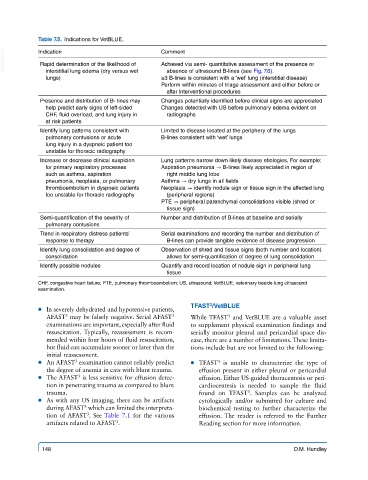
Table 7.5.  Indications for VetBLUE.

  VetBooks.ir  Indication                       Comment
             Rapid determination of the likelihood of
                                                Achieved via semi- quantitative assessment of the presence or
                                                  absence of ultrasound B-lines (see Fig. 7.6).
              interstitial lung edema (dry versus wet
              lungs)                            ≥3 B-lines is consistent with a ‘wet’ lung (interstitial disease)
                                                Perform within minutes of triage assessment and either before or
                                                  after interventional procedures
             Presence and distribution of B- lines may   Changes potentially identified before clinical signs are appreciated
              help predict early signs of left-sided   Changes detected with US before pulmonary edema evident on
              CHF, fluid overload, and lung injury in   radiographs
              at risk patients
             Identify lung patterns consistent with   Limited to disease located at the periphery of the lungs
              pulmonary contusions or acute     B-lines consistent with ‘wet’ lungs
              lung injury in a dyspneic patient too
              unstable for thoracic radiography
             Increase or decrease clinical suspicion   Lung patterns narrow down likely disease etiologies. For example:
              for primary respiratory processes   Aspiration pneumonia → B-lines likely appreciated in region of
              such as asthma, aspiration          right middle lung lobe
              pneumonia, neoplasia, or pulmonary   Asthma → dry lungs in all fields
              thromboembolism in dyspneic patients   Neoplasia → identify nodule sign or tissue sign in the affected lung
              too unstable for thoracic radiography  (peripheral regions)
                                                PTE → peripheral parenchymal consolidations visible (shred or
                                                  tissue sign)
             Semi-quantification of the severity of   Number and distribution of B-lines at baseline and serially
              pulmonary contusions
             Trend in respiratory distress patients’   Serial examinations and recording the number and distribution of
              response to therapy                 B-lines can provide tangible evidence of disease progression
             Identify lung consolidation and degree of   Observation of shred and tissue signs (both number and location)
              consolidation                       allows for semi-quantification of degree of lung consolidation
             Identify possible nodules          Quantify and record location of nodule sign in peripheral lung
                                                  tissue
            CHF, congestive heart failure; PTE, pulmonary thromboembolism; US, ultrasound; VetBLUE, veterinary beside lung ultrasound
            examination.

                                                         TFAST /VetBLUE
                                                              3
             ● ● In severely dehydrated and hypotensive patients,
                                                   3
               AFAST  may be falsely negative. Serial AFAST    While TFAST  and VetBLUE are a valuable asset
                                                                   3
                    3
               examinations are important, especially after fluid   to supplement physical examination findings and
               resuscitation.  Typically, reassessment is recom-  serially monitor pleural and pericardial space dis-
               mended within four hours of fluid resuscitation,   ease, there are a number of limitations. These limita-
               but fluid can accumulate sooner or later than the   tions include but are not limited to the following:
               initial reassessment.
             ● ● An AFAST  examination cannot reliably predict   ● ● TFAST  is unable to characterize the type of
                                                                3
                       3
               the degree of anemia in cats with blunt trauma.  effusion present in either pleural or pericardial
                        3
             ● ● The AFAST  is less sensitive for effusion detec-  effusion. Either US-guided thoracentesis or peri-
               tion in penetrating trauma as compared to blunt   cardiocentesis is needed to sample the fluid
               trauma.                                     found  on  TFAST .  Samples  can  be  analyzed
                                                                         3
             ● ● As with any US imaging, there can be artifacts   cytologically  and/or  submitted  for  culture  and
                          3
               during AFAST  which can limited the interpreta-  biochemical testing to further characterize the
               tion of AFAST . See Table 7.1 for the various   effusion. The reader is referred to the Further
                          3
                                   3
               artifacts related to AFAST .                Reading section for more information.
             148                                                                       D.M. Hundley