
Page 151 - Basic Monitoring in Canine and Feline Emergency Patients
P. 151
Findings of the VetBLUE should be recorded in a access. For the purpose of this chapter, we will discuss
standard fashion for comparison between opera- the dynamic real-time freehand approach.
VetBooks.ir tors and serially over time within one animal. The The US-guided vascular access examination
developers of the VetBLUE have a specific record-
ing protocol (see Further Reading section for more
information). 1. The desired vascular access location is asepti-
cally clipped and cleaned as detailed above, the
entire technique is performed sterilely.
Ultrasound-guided vascular access
2. If attempting central venous catheter placement
In the human literature, there have been descriptions within a large vein, an assistant distends the vein by
of both dynamic real-time and static UST for vascular applying digital pressure proximal to the point of
access. The static technique uses the linear US probe imaging and catheter insertion. If imaging for arte-
to isolate the area of the vascular structure prior to rial catheter access (femoral artery), the area of the
performing a blind venipuncture. The dynamic tech- desired artery is explored with the US without the
nique uses the US to guide the needle into the vessel need for digital occlusion.
in real time. Human medicine describes the use of a 3. The US probe should be manipulated with the
needle guide, but veterinary reports largely use a free- operator’s non-dominant hand while imaging the
hand technique for both arterial and venous vascular desired vessel in either the transverse or longitudinal
(A)
(B)
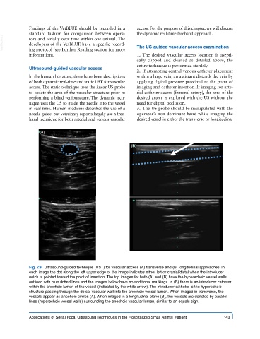
Fig. 7.9. Ultrasound-guided technique (UST) for vascular access (A) transverse and (B) longitudinal approaches. In
each image the dot along the left upper edge of the image indicates either left or cranial/distal when the introducer
notch is pointed toward the point of insertion. The top images for both (A) and (B) have the hyperechoic vessel walls
outlined with blue dotted lines and the images below have no additional markings. In (B) there is an introducer catheter
within the anechoic lumen of the vessel (indicated by the white arrow). The introducer catheter is the hyperechoic
structure passing through the dorsal vascular wall into the anechoic vessel lumen. When imaged in transverse, the
vessels appear as anechoic circles (A). When imaged in a longitudinal plane (B), the vessels are denoted by parallel
lines (hyperechoic vessel walls) surrounding the anechoic vascular lumen, similar to an equals sign.
Applications of Serial Focal Ultrasound Techniques in the Hospitalized Small Animal Patient 143