
Page 530 - Feline diagnostic imaging
P. 530
542 30 Peritoneal Cavity
(a)
(b)
(c) (d)
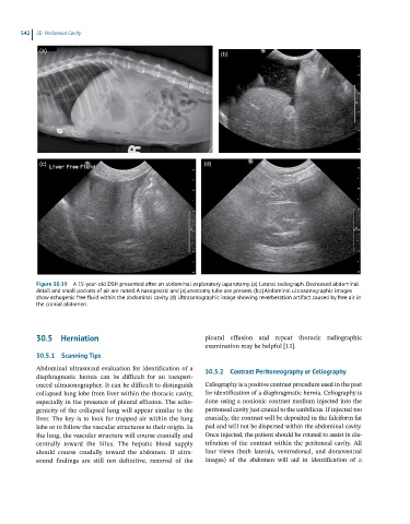
Figure 30.19 A 15-year-old DSH presented after an abdominal exploratory laparotomy. (a) Lateral radiograph. Decreased abdominal
detail and small pockets of air are noted. A nasogastric and jejunostomy tube are present. (b,c) Abdominal ultrasonographic images
show echogenic free fluid within the abdominal cavity. (d) Ultrasonographic image showing reverberation artifact caused by free air in
the cranial abdomen.
30.5 Herniation pleural effusion and repeat thoracic radiographic
examination may be helpful [13].
30.5.1 Scanning Tips
Abdominal ultrasound evaluation for identification of a 30.5.2 Contrast Peritoneography or Celiography
diaphragmatic hernia can be difficult for an inexperi-
enced ultrasonographer. It can be difficult to distinguish Celiography is a positive contrast procedure used in the past
collapsed lung lobe from liver within the thoracic cavity, for identification of a diaphragmatic hernia. Celiography is
especially in the presence of pleural effusion. The echo- done using a nonionic contrast medium injected into the
genicity of the collapsed lung will appear similar to the peritoneal cavity just cranial to the umbilicus. If injected too
liver. The key is to look for trapped air within the lung cranially, the contrast will be deposited in the falciform fat
lobe or to follow the vascular structures to their origin. In pad and will not be dispersed within the abdominal cavity.
the lung, the vascular structure will course cranially and Once injected, the patient should be rotated to assist in dis-
centrally toward the hilus. The hepatic blood supply tribution of the contrast within the peritoneal cavity. All
should course caudally toward the abdomen. If ultra- four views (both laterals, ventrodorsal, and dorsoventral
sound findings are still not definitive, removal of the images) of the abdomen will aid in identification of a