
Page 535 - Feline diagnostic imaging
P. 535
30.5 Herniation 547
(b)
(a)
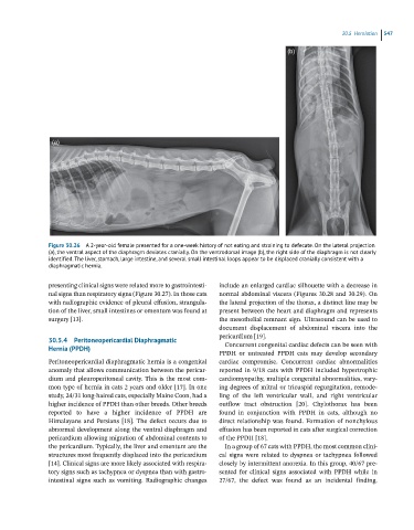
Figure 30.26 A 2-year-old female presented for a one-week history of not eating and straining to defecate. On the lateral projection
(a), the ventral aspect of the diaphragm deviates cranially. On the ventrodorsal image (b), the right side of the diaphragm is not clearly
identified. The liver, stomach, large intestine, and several small intestinal loops appear to be displaced cranially consistent with a
diaphragmatic hernia.
presenting clinical signs were related more to gastrointesti- include an enlarged cardiac silhouette with a decrease in
nal signs than respiratory signs (Figure 30.27). In those cats normal abdominal viscera (Figures 30.28 and 30.29). On
with radiographic evidence of pleural effusion, strangula- the lateral projection of the thorax, a distinct line may be
tion of the liver, small intestines or omentum was found at present between the heart and diaphragm and represents
surgery [13]. the mesothelial remnant sign. Ultrasound can be used to
document displacement of abdominal viscera into the
pericardium [19].
30.5.4 Peritoneopericardial Diaphragmatic
Hernia (PPDH) Concurrent congenital cardiac defects can be seen with
PPDH or untreated PPDH cats may develop secondary
Peritoneopericardial diaphragmatic hernia is a congenital cardiac compromise. Concurrent cardiac abnormalities
anomaly that allows communication between the pericar- reported in 9/18 cats with PPDH included hypertrophic
dium and pleuroperitoneal cavity. This is the most com- cardiomyopathy, multiple congenital abnormalities, vary-
mon type of hernia in cats 2 years and older [17]. In one ing degrees of mitral or tricuspid regurgitation, remode-
study, 24/31 long‐haired cats, especially Maine Coon, had a ling of the left ventricular wall, and right ventricular
higher incidence of PPDH than other breeds. Other breeds outflow tract obstruction [20]. Chylothorax has been
reported to have a higher incidence of PPDH are found in conjunction with PPDH in cats, although no
Himalayans and Persians [18]. The defect occurs due to direct relationship was found. Formation of nonchylous
abnormal development along the ventral diaphragm and effusion has been reported in cats after surgical correction
pericardium allowing migration of abdominal contents to of the PPDH [18].
the pericardium. Typically, the liver and omentum are the In a group of 67 cats with PPDH, the most common clini-
structures most frequently displaced into the pericardium cal signs were related to dyspnea or tachypnea followed
[14]. Clinical signs are more likely associated with respira- closely by intermittent anorexia. In this group, 40/67 pre-
tory signs such as tachypnea or dyspnea than with gastro- sented for clinical signs associated with PPDH while in
intestinal signs such as vomiting. Radiographic changes 27/67, the defect was found as an incidental finding.