
Page 536 - Feline diagnostic imaging
P. 536
(a)
(b)
(c)
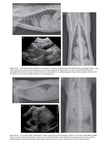
Figure 30.27 A 10-year-old DSH presented for evaluation of a caudal mammary mass. (a) A lateral thoracic radiograph shows a mass
in the caudal thorax of soft tissue and fat opacity. The ventral border of the diaphragm is not well visualized. (b) Ventrodorsal
radiograph. Decreased liver volume is noted within the cranial abdomen. (c) Ultrasonographic image shows a portion of liver within
the thoracic cavity. A chronic diaphragmatic hernia was diagnosed.
(a) (b)
(c)
Figure 30.28 A 6-month-old DLH presented for chronic cough. (a) Lateral radiographic projection. The cardiac silhouette is greatly
enlarged. (b) Ventrodorsal radiographic projection. The cardiac silhouette extends laterally to occupy the entire thoracic cavity. (c)
Ultrasonographic image shows displacement of small intestinal loops, liver, and a cystic structure within the pericardium.