
Page 532 - Feline diagnostic imaging
P. 532
(a)
(b)
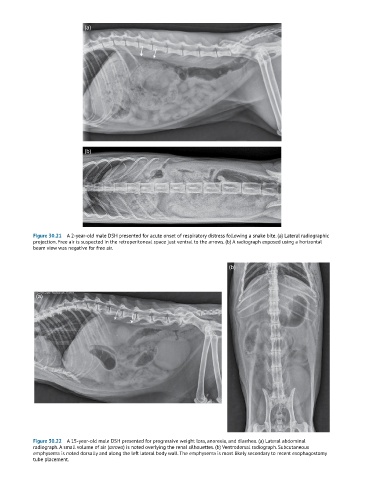
Figure 30.21 A 2-year-old male DSH presented for acute onset of respiratory distress following a snake bite. (a) Lateral radiographic
projection. Free air is suspected in the retroperitoneal space just ventral to the arrows. (b) A radiograph exposed using a horizontal
beam view was negative for free air.
(b)
(a)
Figure 30.22 A 15-year-old male DSH presented for progressive weight loss, anorexia, and diarrhea. (a) Lateral abdominal
radiograph. A small volume of air (arrows) is noted overlying the renal silhouettes. (b) Ventrodorsal radiograph. Subcutaneous
emphysema is noted dorsally and along the left lateral body wall. The emphysema is most likely secondary to recent esophagostomy
tube placement.