
Page 537 - Feline diagnostic imaging
P. 537
30.5 Herniation 549
(b)
(a)
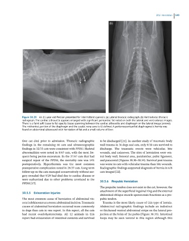
Figure 30.29 An 11-year-old Persian presented for intermittent cyanosis. (a) Lateral thoracic radiograph. (b) Ventrodorsal thoracic
radiograph. The cardiac silhouette appears enlarged with significant pericardial fat noted on both the lateral and ventrodorsal images.
There is a faint soft tissue to fat opacity tissue spanning between the cardiac silhouette and diaphragm on the lateral image (arrows).
The midventral portion of the diaphragm and the caudal vena cava is ill defined. A peritoneopericardial diaphragmatic hernia was
found on abdominal ultrasound with herniation of fat and a small volume of liver.
One cat died prior to admission. Thoracic radiographic to be discharged [21]. In another study of traumatic body
findings in the remaining 66 cats and ultrasonographic wall trauma in 36 dogs and cats, only 8/10 cats survived to
findings in 32/33 cats were consistent with PPDH. Skeletal discharge. The traumatic events were vehicular, bite
abnormalities were noted in 8/67 cats, with the most fre- wounds, and unknown. The sites of herniation were ven-
quent being pectus excavatum. In the 37/67 cats that had tral body wall, femoral area, paralumbar, pubic ligament,
surgical repair of the PPDH, the mortality rate was 14% and paracostal (Figures 30.30–30.34). Survival post trauma
postoperatively. Hyperthermia was the most common was worse in cats with vehicular trauma than bite wounds.
postoperative complication noted in 29/37 cats. Long‐term Radiographic findings supported diagnosis of hernia in 6/8
follow‐up on the cats managed conservatively without sur- cats imaged [22].
gery revealed that 9/29 had died due to cardiac disease or
were euthanized due to other problems unrelated to the
PPDH [17]. 30.5.6 Prepubic Herniation
The prepubic tendon does not exist in the cat; however, the
attachment of the superficial inguinal ring and the external
30.5.5 Evisceration Injuries
abdominal oblique muscle aponeurosis functions as a pre-
The most common cause of herniation of abdominal vis- pubic tendon.
cera is dehiscence at a recent abdominal incision. Traumatic Trauma is the most likely cause of this type of hernia.
causes of abdominal herniation occurred more commonly Abdominal radiographic findings include an indistinct
in dogs than cats in one report. In this report, all the cats or thickened ventral abdominal stripe on the lateral pro -
had recent ovariohysterectomy. All 12 animals in this jection at the brim of the pubis (Figure 30.35). Intestinal
report had evisceration of intestinal contents and survived loops may be seen ventral to this region although this