
Page 533 - Feline diagnostic imaging
P. 533
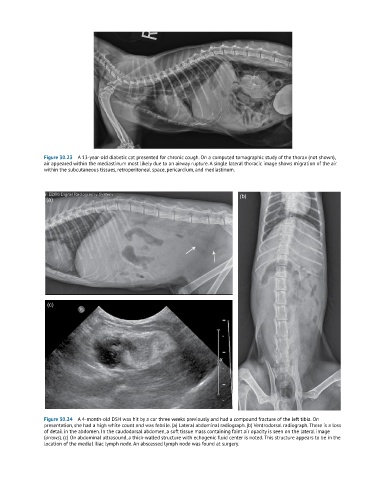
Figure 30.23  A 13-year-old diabetic cat presented for chronic cough. On a computed tomographic study of the thorax (not shown),
               air appeared within the mediastinum most likely due to an airway rupture. A single lateral thoracic image shows migration of the air
               within the subcutaneous tissues, retroperitoneal space, pericardium, and mediastinum.


                                                                               (b)
                (a)


                (c)


               Figure 30.24  A 4-month-old DSH was hit by a car three weeks previously and had a compound fracture of the left tibia. On
               presentation, she had a high white count and was febrile. (a) Lateral abdominal radiograph. (b) Ventrodorsal radiograph. There is a loss
               of detail in the abdomen. In the caudodorsal abdomen, a soft tissue mass containing faint air opacity is seen on the lateral image
               (arrows). (c) On abdominal ultrasound, a thick-walled structure with echogenic fluid center is noted. This structure appears to be in the
               location of the medial iliac lymph node. An abscessed lymph node was found at surgery.