
Page 538 - Feline diagnostic imaging
P. 538
550 30 Peritoneal Cavity
(b)
(a)
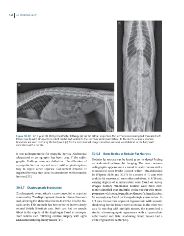
Figure 30.30 A 16-year-old DSH presented for lethargy. (a) On the lateral projection, the sternum was malaligned. Increased soft
tissue opacity with air opacity is noted caudal and ventral to the sternum. Ventral and lateral to the mid to caudal abdomen,
intestines are seen overlying the body wall. (b) On the ventrodorsal image, intestines are seen caudolateral to the body wall
consistent with a hernia.
is not pathognomonic for prepubic hernia. Abdominal 30.5.8 Bates Bodies or Nodular Fat Necrosis
ultrasound or celiography has been used if the radio -
graphic findings were not definitive. Identification of Nodular fat necrosis can be found as an incidental finding
on abdominal radiographic imaging. The most common
a prepubic hernia may not occur until surgical explora-
tory to repair other injuries. Concurrent femoral or radiographic appearance is a round to oval structure with a
mineralized outer border located within intraabdominal
inguinal hernias may occur in association with prepubic
hernias [23]. fat (Figures 30.36 and 30.37). In a report of 10 cats with
nodular fat necrosis, all were older and obese. In 9/10 cats,
varying degrees of mineralization were found on survey
images. Solitary mineralized nodules were more com-
30.5.7 Diaphragmatic Eventration
monly identified than multiple. In the one cat with multi-
Diaphragmatic eventration is a rare congenital or acquired ple masses with no radiographic evidence of mineralization,
abnormality. The diaphragmatic tissue is thinner than nor- fat necrosis was found on histopathologic examination. In
mal, allowing the abdominal viscera to extend into the tho- 1/3 cats, fat necrosis appeared hyperechoic with acoustic
racic cavity. This anomaly has been reported in two closely shadowing but the lesions were not found in the other two
related British Shorthair cats. Both cats had no muscle cats. In one dog with multiple masses, the masses had a
fibers in the cupula of the diaphragm found at necropsy. similar ultrasonographic appearance with a hyperechoic
Both kittens died following elective surgery with signs outer border and distal shadowing. Some masses had a
associated with respiratory failure [24]. visible hypoechoic center [25].