
Page 541 - Feline diagnostic imaging
P. 541
(b)
(a)
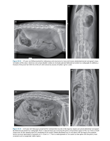
Figure 30.34 A 9-year-old DSH presented for listlessness and reluctance to move. (a) A lateral abdominal lateral radiograph shows
herniation of the small intestines and urinary bladder ventral to the body wall. (b) Ventrodorsal abdominal radiograph. In addition, a
fracture of the proximal 11th rib on the left and transverse process of lumbar spine is seen.
(b)
(a)
Figure 30.35 A 14-year-old Himalayan presented for multiple bite wounds in the inguinal region. (a) Lateral abdominal radiograph.
(b) Ventrodorsal abdominal radiograph. There is subcutaneous gas overlying the pelvis and proximal aspect of the rear limbs on both
projections. On the lateral projection, thickening of the caudal ventral abdominal wall is consistent with damage to the prepubic
tendon (arrows). Spondylosis is present at L5–6 and L6–7. There is malalignment of the caudal lumbar spine with the pelvis, most
consistent with a congenital malformation.