
Page 539 - Feline diagnostic imaging
P. 539
30.5 Herniation 551
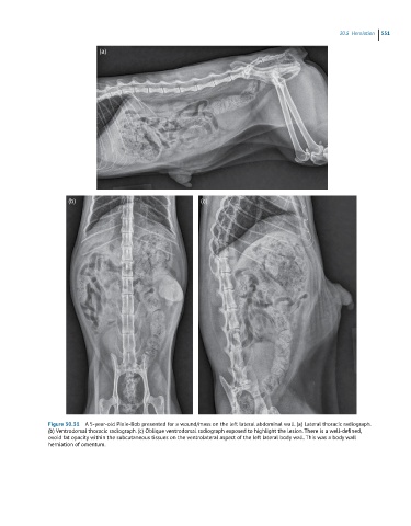
(a)
(b) (c)
Figure 30.31 A 5-year-old Pixie-Bob presented for a wound/mass on the left lateral abdominal wall. (a) Lateral thoracic radiograph.
(b) Ventrodorsal thoracic radiograph. (c) Oblique ventrodorsal radiograph exposed to highlight the lesion. There is a well-defined,
ovoid fat opacity within the subcutaneous tissues on the ventrolateral aspect of the left lateral body wall. This was a body wall
herniation of omentum.