
Page 540 - Feline diagnostic imaging
P. 540
(b)
(a)
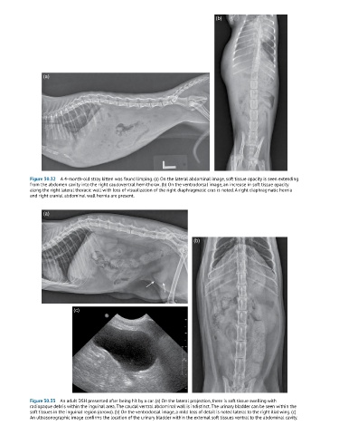
Figure 30.32 A 4-month-old stray kitten was found limping. (a) On the lateral abdominal image, soft tissue opacity is seen extending
from the abdomen cavity into the right caudoventral hemithorax. (b) On the ventrodorsal image, an increase in soft tissue opacity
along the right lateral thoracic wall with loss of visualization of the right diaphragmatic crus is noted. A right diaphragmatic hernia
and right cranial abdominal wall hernia are present.
(a)
(b)
(c)
Figure 30.33 An adult DSH presented after being hit by a car. (a) On the lateral projection, there is soft tissue swelling with
radiopaque debris within the inguinal area. The caudal ventral abdominal wall is indistinct. The urinary bladder can be seen within the
soft tissues in the inguinal region (arrows). (b) On the ventrodorsal image, a mild loss of detail is noted lateral to the right ilial wing. (c)
An ultrasonographic image confirms the location of the urinary bladder within the external soft tissues ventral to the abdominal cavity.