
Page 542 - Feline diagnostic imaging
P. 542
(b)
(a)
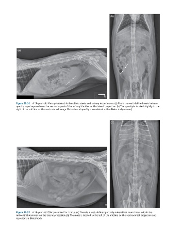
Figure 30.36 A 14-year-old Manx presented for hindlimb ataxia and urinary incontinence. (a) There is a well-defined ovoid mineral
opacity superimposed over the ventral aspect of the urinary bladder on the lateral projection. (b) The opacity is located slightly to the
right of the midline on the ventrodorsal image. This mineral opacity is consistent with a Bates body (arrows).
(b)
(a)
Figure 30.37 A 13-year-old DSH presented for icterus. (a) There is a well-defined partially mineralized round mass within the
midventral abdomen on the lateral projection. (b) The mass is located to the left of the midline on the ventrodorsal projection and
represents a Bates body.