
Page 546 - Feline diagnostic imaging
P. 546
31.2 Diseisi AAsecDing ctse Dii 559
(b)
(a)
(c)
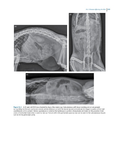
Figure 31.2 A 19-year-old DSH was attacked by dogs a few weeks ago. Subcutaneous soft tissue swelling and air are present
surrounding the thoracic cavity and cranial ventral abdomen on both the lateral (a) and ventrodorsal (b) images. Luxation of the right
coxofemoral joint is present. Ventral spondylosis is noted at the lumbosacral junction. Osteophytes are noted on the proximal tibial
crests. A horizontal beam view is taken to rule out free air within the peritoneal space (c). Gas can be seen in the subcutaneous tissues
but not in the peritoneal cavity.