
Page 549 - Feline diagnostic imaging
P. 549
562 31 Body Wall
(b)
(a)
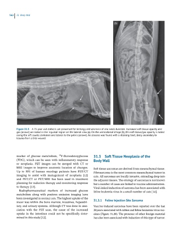
Figure 31.5 A 15-year-old diabetic cat presented for lethargy and anorexia of one week duration. Increased soft tissue opacity and
gas (arrows) are noted in the inguinal region on the lateral view (a). On the ventrodorsal image (b), this soft tissue/gas opacity is noted
along the left caudal abdomen and lateral to the pelvis (arrows). An abscess was found with a draining tract, likely secondary to
trauma from a bite wound.
18
marker of glucose metabolism, F-fluroodexoyglucose 31.3 Soft Tissue Neoplasia of the
(FDG), which can be seen with inflammatory response Body Wall
or neoplasia. PET images can be merged with CT or
MRI images to improve anatomic location of changes. Soft tissue sarcomas are derived from mesenchymal tissue.
Up to 50% of human oncology patients have PET/CT Fibrosarcoma is the most common mesenchymal tumor in
imaging to assist with management of neoplasia [12] cats. All sarcomas are locally invasive, extending deep into
and PET/CT or PET/MRI has been used in treatment the adjacent tissues. The etiology of sarcomas is not known
planning for radiation therapy and monitoring response but a number of cases are linked to vaccine administration.
to therapy [13]. Viral-linked induction of sarcoma has been associated with
Radiopharmaceutical markers of increased glucose feline leukemia virus in a small number of cats [14].
metabolism along with positron emission imaging have
been investigated in normal cats. The highest uptake of the
tracer was within the bone marrow, intestines, hepatobil- 31.3.1 Feline Injection Site Sarcoma
iary, and urinary systems. Although CT was done in asso- Vaccine-induced sarcomas have been reported over the last
ciation with the PET scan, the cause of the increased 20 years associated with rabies and feline leukemia virus vac-
uptake in the intestines could not be specifically deter- cines (Figure 31.20). The presence of other foreign material
mined in this study [12]. has also been associated with induction of this type of tumor.