
Page 554 - Feline diagnostic imaging
P. 554
31.3 SAc Diiss sSopeiDe SAects Sody epp 567
(b)
(a)
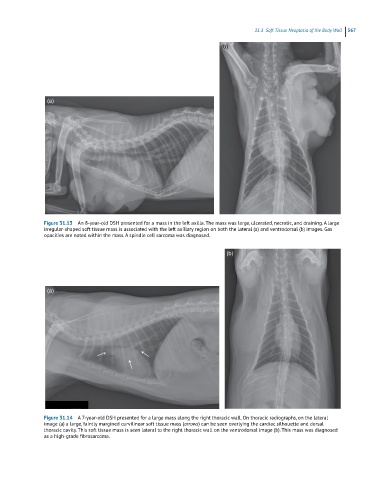
Figure 31.13 An 8-year-old DSH presented for a mass in the left axilla. The mass was large, ulcerated, necrotic, and draining. A large
irregular-shaped soft tissue mass is associated with the left axillary region on both the lateral (a) and ventrodorsal (b) images. Gas
opacities are noted within the mass. A spindle cell sarcoma was diagnosed.
(b)
(a)
Figure 31.14 A 7-year-old DSH presented for a large mass along the right thoracic wall. On thoracic radiographs, on the lateral
image (a) a large, faintly margined curvilinear soft tissue mass (arrows) can be seen overlying the cardiac silhouette and dorsal
thoracic cavity. This soft tissue mass is seen lateral to the right thoracic wall on the ventrodorsal image (b). This mass was diagnosed
as a high-grade fibrosarcoma.