
Page 553 - Feline diagnostic imaging
P. 553
566 31 Body Wall
(b)
(a)
(c) (d)
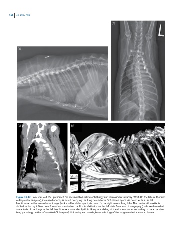
Figure 31.12 A 6-year-old DSH presented for one month duration of lethargy and increased respiratory effort. On the lateral thoracic
radiographic image (a), increased opacity is noted overlying the lung parenchyma. Soft tissue opacity is noted within the left
hemithorax on the ventrodorsal image (b). A small nodular opacity is noted in the right cranial lung lobe. The cardiac silhouette is
shifted to the right. New bone formation is noted on the first to sixth ribs on the left side. Computed tomography (c) showed rounded
atelectasis of the lungs in the left hemithorax surrounded by fluid. Bony remodeling of the ribs was noted secondary to the extensive
lung pathology on the reformatted CT image (d). Following euthanasia, histopathology of the lung revealed adenocarcinoma.